Return to Case List with Diagnoses or Case List as Unknowns

PET/CT IMAGING FOR EPILEPTIC LOCALIZATION
Authored By: Peter Phan and Akash Sharma.
Patient: 17 year old female
History:

The patient is a 17-year-old girl with intractable dominant hemisphere motor seizures and recent breakthrough episodes. Video EEG findings suggest that the seizures may be emanating from the left frontal lobe.  However, a recent MRI was unremarkable for an epileptic focus and was read out as normal.

Image Size:[small][as-submitted]

Fig. 1
Axial PET images

Fig. 2
Coronal PET images

Fig. 3
PET and corresponding T1 axial images

Fig. 4
T2 MRI images
Image Size:[small][as-submitted]

Findings:

On the PET images, two hypermetabolic foci are demonstrated within the left frontal cortex.  Although the patient's MRI was described as normal, direct correlation with the PET images revealed corresponding abnormal focal cortical thickening.

DDx:

Focal cortical dysplasia

Diagnosis:

Focal cortical dysplasia

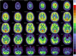
General Discussion:

RADIOPHARMACEUTICAL: 8.6 mCi F-18 Fluorodeoxyglucose i.v.

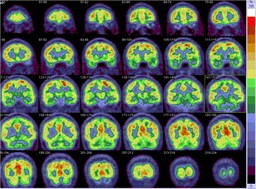
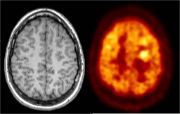
For the purpose of epileptic localization, PET imaging is usually done as an interictal evaluation.  PET imaging for ictal evaluation are not performed because of the underlying technical challenges.   Positive findings for interictal exams are generally hypometabolic, typically related to mesial temporal sclerosis.   In this patient, who has a recent MRI that was unremarkable for any CNS malignancy, the unexpected findings of two hypermetabolic foci may reflect an ictal phase imaging rather than interictal.  The patient was heavily sedated and medicated during the exam, which would explain the lack of any physical manifestation of her ongoing seizures during the uptake phase of the study. 

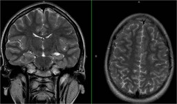
Therefore, in combination with the subtle MRI finding and concordance of the video EEG, this is likely to represent focal cortical dysplasia and the patient's seizure focus.

The patient underwent surgical resection for these abnormal foci, which was revealed to be focal cortical dyplasia on pathology.  Subsequently, she was seizure free.

Comments:
No comments posted.
Additional Details:

Case Number: 122053Owner(s): Peter Phan and Akash SharmaLast Updated: 12-07-2011
Anatomy: Cranium and Contents   Pathology: Congenital
Modality: PETAccess Level: Readable by all users, writable by NucMed Certifiers
Keywords: pbnmACR: 10000.15000

Case has been viewed 47 times.
Certified by Akash Sharma on 03-10-2010

The reader is fully responsible for confirming the accuracy of this content.
Text and images may be copyrighted by the case author or institution.