Return to Case List with Diagnoses or Case List as Unknowns

LEFT RENAL ARTERY STENOSIS
Authored By: Xiaoni Hong and Barry Siegel, Prof of Radiology.
Patient: 2 year 1 month 14 day old male
History: 2-year-old boy admitted with hypertensive emergency.
Image Size:[small][as-submitted]

Fig. 1
Posterior radionuclide angiogram demonstrates decreased perfusion to the smaller left kidney

Fig. 2
Sequential renal scintigrapy images show decreased function of the smaller left kidney, with progressively increasing renal parenchymal uptake of tracer, but no excretion. In light of the clinical history, the findings are most consistent with high-grade left renal artery stenosis. Normal right renal function with no evidence of obstruction.

Fig. 3
Post-voiding image.

Fig. 4
CT angiograhy reveals 6 mm focal stenosis of the left renal artery in its proximal portion. Distal to the stenosis the artery caliber increases to 4 mm in diameter.There is decreased size and a delayed nephrogram of the left kidney.
Image Size:[small][as-submitted]

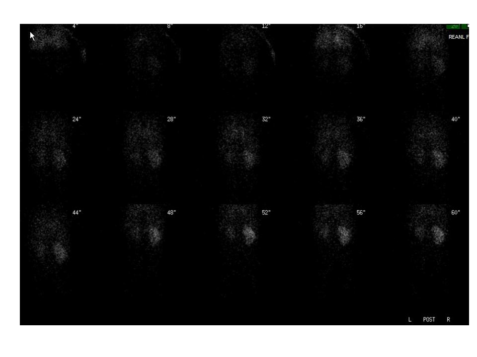
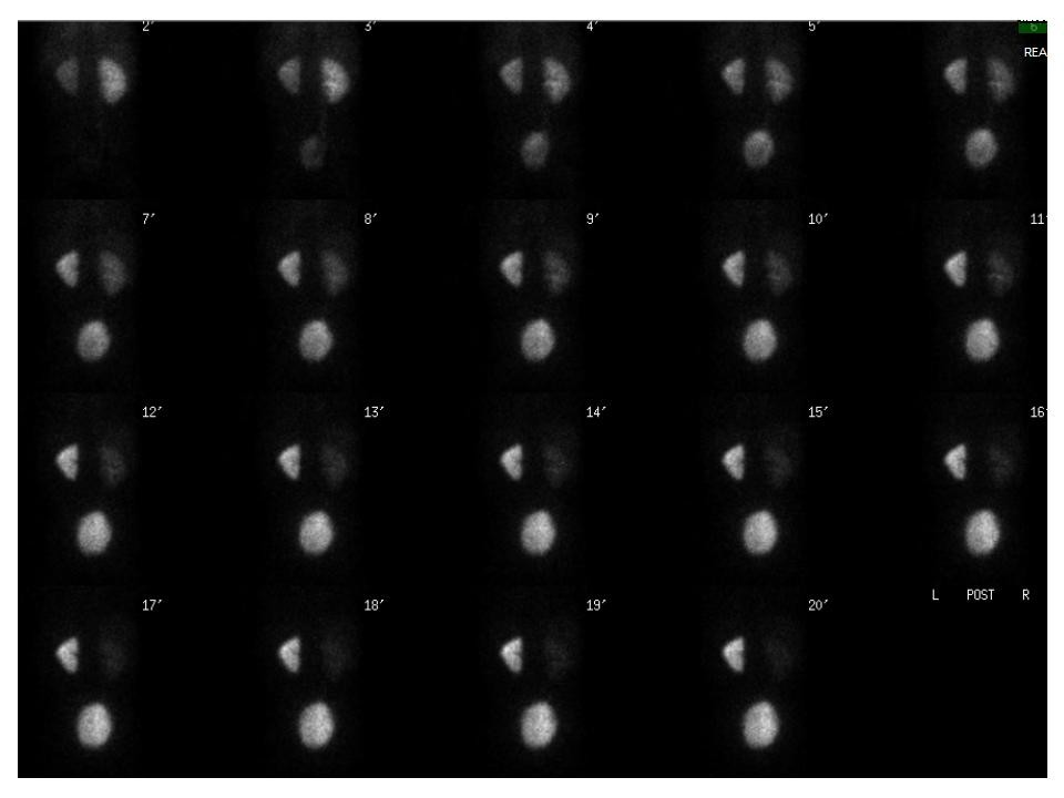
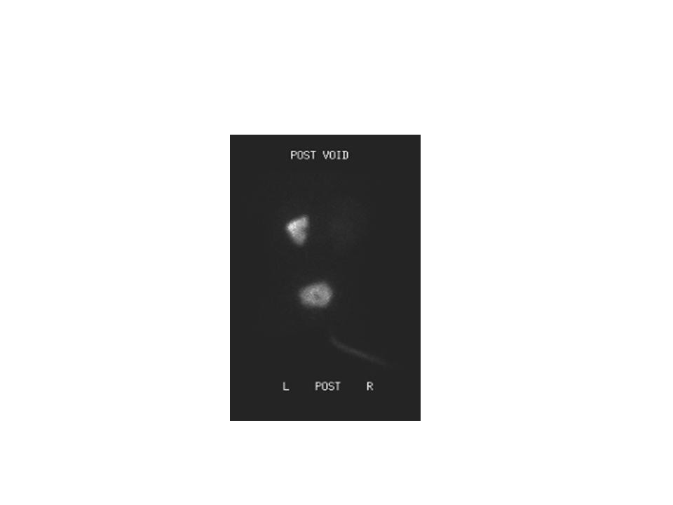
Findings:

Renal scintigraphy demonstrated decreased perfusion and function of the smaller left kidney, with progressively increasing renal parenchymal uptake of tracer, but no excretion. In light of the clinical history, the findings are most consistent with high-grade left renal artery stenosis. There is normal right renal function with no evidence of obstruction.

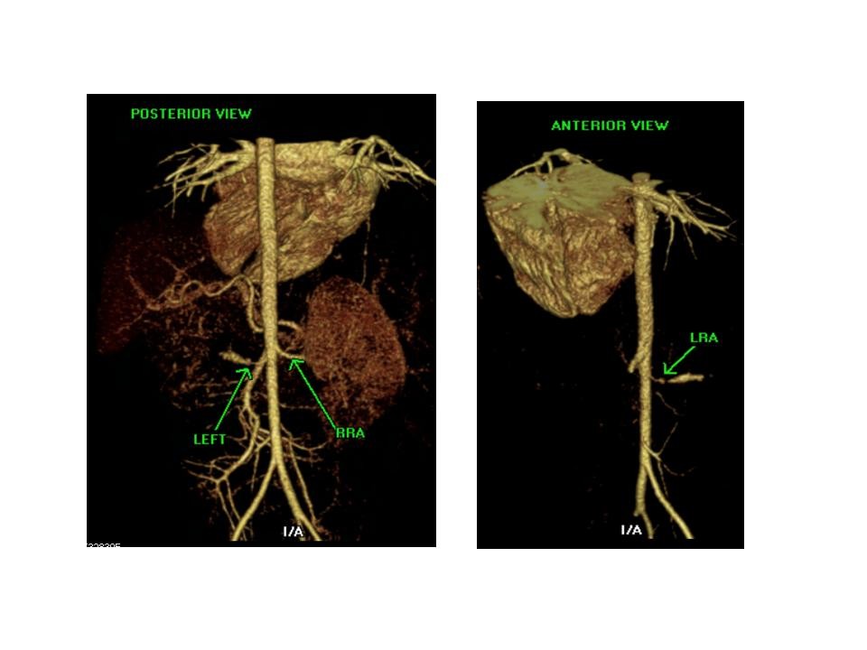
CT angiograhy deminstrated a 6 mm focal stenosis of the left renal artery in its proximal portion. Distal to the stenosis the artery caliber increases to 4 mm in diameter.
There is decreased size and a delayed nephrogram of the left kidney, consistent with focal left renal artery stenosis.


DDx:    Unilateral renal artery stenosis
   Unilateral ATN
   Unilateral renal vein thrombosis
   Unilateral high-grade obstruction
Diagnosis: Left renal artery stenosis
General Discussion:

Long history: 2-year-old boy admitted with hypertensive emergency. At another hospital, ultrasonography demonstrated discrepant kidney size, with the left kidney measuring approximately 2 cm less in length than the right kidney. CT angiogram showed a left renal artery stenosis. He is currently being treated with the nicardipine and nitroprusside. He has not received an ACE inhibitor or an angiotensin receptor blocker. Serum creatinine is 0.3 mg/dL. Evaluate relative renal function.

Radiopharmaceutical: 1.0 mCi Tc-99m MAG3 i.v.

Follow up: Patient underwent left retroperitoneal exploration with ligation of left renal artery and reimplantation into the aorta. Pathology reported ARTERY WITH INTIMAL FIBROMUSCULAR TISSUE, CONSISTENT WITH STENOSIS.

Specific Discussion: Renovascular hypertension (RVH) is the most common cause of surgically correctable hypertension in children. In North America and Western Europe, fibromuscular dysplasia is the most common etiology of pediatric RVH, whereas in Asia and Africa, aortoarteritis is the most common cause. The fibromuscular dysplasia that is observed in the pediatric population does not demonstrate the classic “string of beads” pattern seen in adults. Instead, the stenosis generally appears as discrete narrowing on angiography.

ACE-inhibited renal scintigraphy is the most specific scintigraphic method for evalauting suspected RVH.  In this patient with documented renal artery stenosis, the scintigraphic findings without ACE inhibition are sufficiently characteristic to permit a confident diagnsosis of RVH. 
References:

1. Aryce D, Beseth MD. Renal artery aneurysm secondary to fibromuscular dysplasia in a young patient. Ann Vasc Surg. 2005; 19: 605-608.

2. Ziessman HA. The Requisites: Nuclear Medicine. 3rd edition; p230-234.

Comments:
No comments posted.
Additional Details:

Case Number: 96458Owner(s): Xiaoni Hong and Barry Siegel, Prof of RadiologyLast Updated: 02-07-2013
Anatomy: Genitourinary (GU)   Pathology: Vascular
Modality: Nuc MedAccess Level: Readable by all users, writable by NucMed Certifiers
Keywords: rsnmACR: 80000.12170

Case has been viewed 48 times.
Certified by Barry Siegel on 06-13-2009

The reader is fully responsible for confirming the accuracy of this content.
Text and images may be copyrighted by the case author or institution.