Return to Case List with Diagnoses or Case List as Unknowns

HEPATIC SUBCAPSULAR HEMATOMA
Authored By: tdeshm01 and Barry Siegel, Prof of Radiology.
Patient: 22 year old female
History: 22-year-old woman with fever, shortness of breath and right upper quadrant abdominal pain.
Image Size:[small][as-submitted]

Fig. 1
Anterior images for 1 min

Fig. 2
Posterior images for 1 min

Fig. 3
Anterior images for 1 hour

Fig. 4
Posterior images for 1 hour

Fig. 5
Anterior and Posterior images after 1.5 hours delay

Fig. 6
Anterior and Posterior images after 4 hours delay

Fig. 7
CECT Abdomen
Image Size:[small][as-submitted]

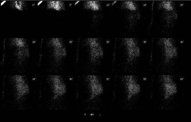
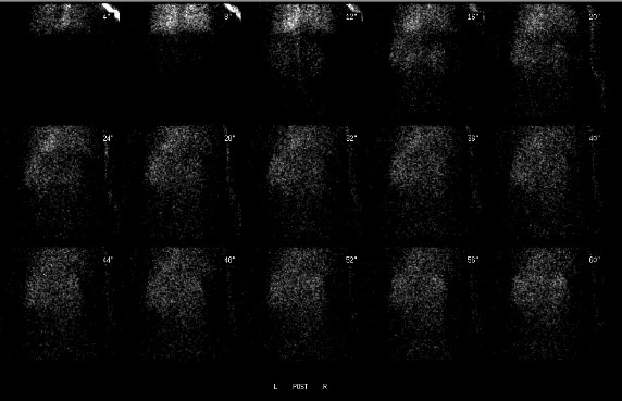
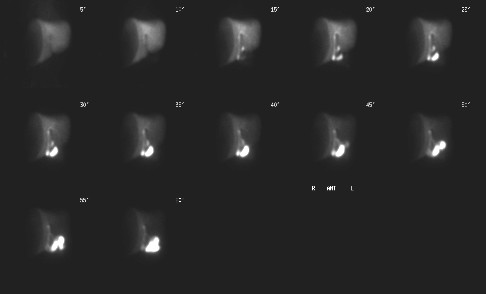
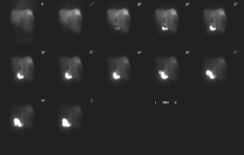
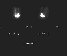
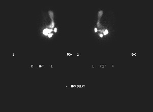
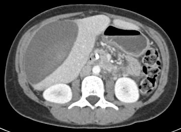
Findings:

RADIOPHARMACEUTICAL: 3.3 mCi Tc-99m mebrofenin i.v.

FINDINGS: The radionuclide angiogram and sequential images demonstrate a large photopenic region with a concave medial border involving most of the right lobe of the liver. There is prompt, uniform accumulation of the tracer by the remainder of the liver. No activity is seen to accumulate in the photopenic region (thus excluding a biloma).

There is normal filling of the intrahepatic ducts and normal excretion of the tracer into the duodenum via the stented common bile duct. Early in the imaging sequence, there appears to be reflux of tracer into the antrum of the stomach. The 4-hour delayed images show filling of the entire stomach with tracer.

The tracer excreted into the duodenum moves very slowly within the bowel lumen, and did not progress beyond the proximal jejunum on the 4-hour images.This is most consistent with ileus.

DDx:

1. Hepatic neoplasm

2. Hepatic abscess

Diagnosis:

1. Large right hepatic subcapsular hematoma.

2. Common bile duct stent in place without evidence of bile leak.

3. Probable ileus.

4. Duodenogastric reflux.

General Discussion:

FULL PATIENT HISTORY: 22-year-old woman with fever, shortness of breath, abdominal pain and abnormal liver function tests 2 weeks after laparoscopic cholecystectomy.  An abdominal CT scan demonstrated a right hepatic subcapsular fluid collection. Subsequently, ERCP was performed with sphincterotomy and common bile duct stent placement. There was no evidence of bile leak. On the next day, hepatobiliary scintigraphy was requested for further assessment of a possible bile leak.

DISCUSSION: Tc-99m IDA radiopharmaceuticals are concentrated by the hepatocytes and secreted into the bile canaliculi and then into the biliary radicals, bile duct, gall bladder and small intestine. Common indications for hepatobiliary scintigraphy include diagnosis of acute cholecystitis, documentation mof biliary tract patency, identification of biliary leaks and differentiation of biliary atresia from neonatal hepatitis in neonates. Less common indications are evaluation of biliary dyskinesia and sphincter of Oddi dysfunction.

In this post cholecystectomy patient, the ERCP and HIDA scan were performed to evaluate whether the hepatic subcapsular fluid collection demonstrated on the abdominal CT scan was secondary to a bile leak. However, no biliary leak was identified and the appearance of the fluid collection was most consistent with a supcapsular hematoma secondary to the recent surgery.

Comments:
No comments posted.
Additional Details:

Case Number: 198306Owner(s): tdeshm01 and Barry Siegel, Prof of RadiologyLast Updated: 02-07-2013
Anatomy: Gastrointestinal (GI)   Pathology: Iatrogenic
Modality: CT, Nuc MedAccess Level: Readable by all users, writable by NucMed Certifiers
Keywords: hsnm

Case has been viewed 40 times.
Certified by Barry Siegel on 01-02-2010

The reader is fully responsible for confirming the accuracy of this content.
Text and images may be copyrighted by the case author or institution.