Return to Case List with Diagnoses or Case List as Unknowns

HEPATIC CARCINOID
Authored By: Keith Fischer, Shane Inoue and Jerold Wallis, Assoc Prof of Radiology.
Patient: 54 year old male
History: 54-year-old male with imaging requested as part of a pre-transplant evaluation.
Image Size:[small][as-submitted]

Fig. 1
Whole body scan

Fig. 2
Whole body scan

Fig. 3
Liver CT

Fig. 4
Liver MR (T2 and Post-contrast)
Image Size:[small][as-submitted]

Findings: Bone Scan: Delayed whole-body scintigrams were obtained. There is normal distribution of activity throughout the skeleton. Mild diffuse increased uptake is seen in the abdomen consistent with ascites.

Octreoscan: There is markedly increased In-111 pentetreotide activity in the liver, most pronounced in the right hepatic lobe, but involving the entire liver.

CT:  Hepatomegaly with vague low attenuation lesions.

MRI:   Marked nodularity throughout the liver which is markedly enlarged, suggesting tumor involvement.
Diagnosis: Hepatic Carcinoid
General Discussion: 54-year-old male with symptomatic carcinoid tumor of the liver with resultant liver failure and portal hypertension. This scan is requested as part of a preliver transplant evaluation.

There is markedly increased In-111 pentetreotide activity in the liver, to a degree that makes it difficult to identify this study as an In-111 Octreotide study.  While some of this uptake may reflect poor liver clearance of tracer, the majority of uptake is likely due to tumor. The liver is enlarged, and appears to be nearly completely replaced by tumor.
Normally, spleen and kidneys are more intense than liver on an Octreotide study, whereas in this case the liver is much more intense.

The intense liver uptake limits evaluation of potential uptake within adjacent soft tissues and lymph nodes. There is physiologic uptake within spleen and the kidneys.

CT and MR again show hepatomegaly, with nodularity from the tumor involvement.
Comments:
No comments posted.
Additional Details:

Case Number: 132539Owner(s): Keith Fischer, Shane Inoue and Jerold Wallis, Assoc Prof of RadiologyLast Updated: 02-07-2013
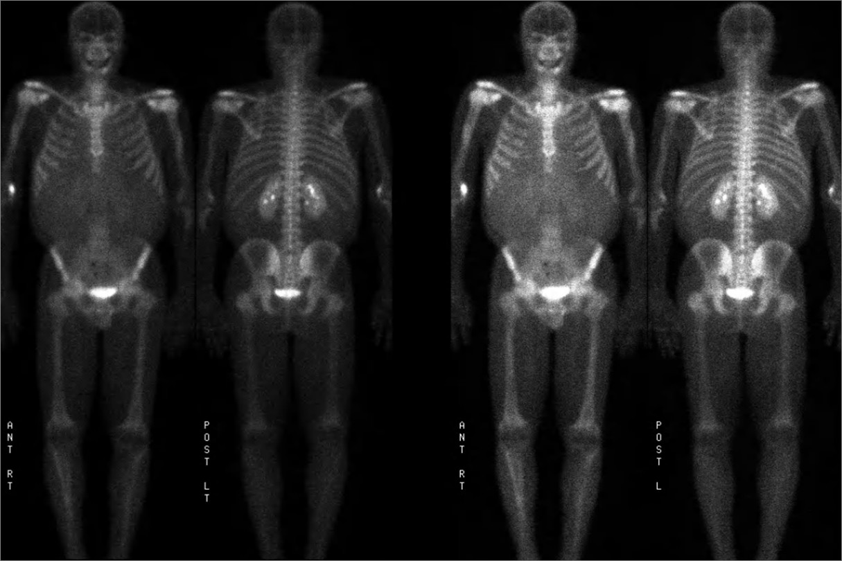
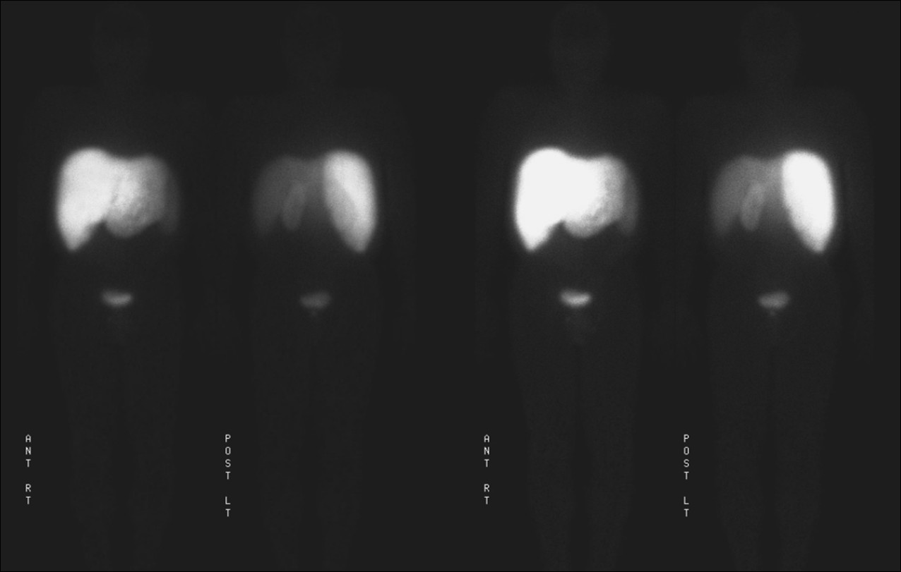
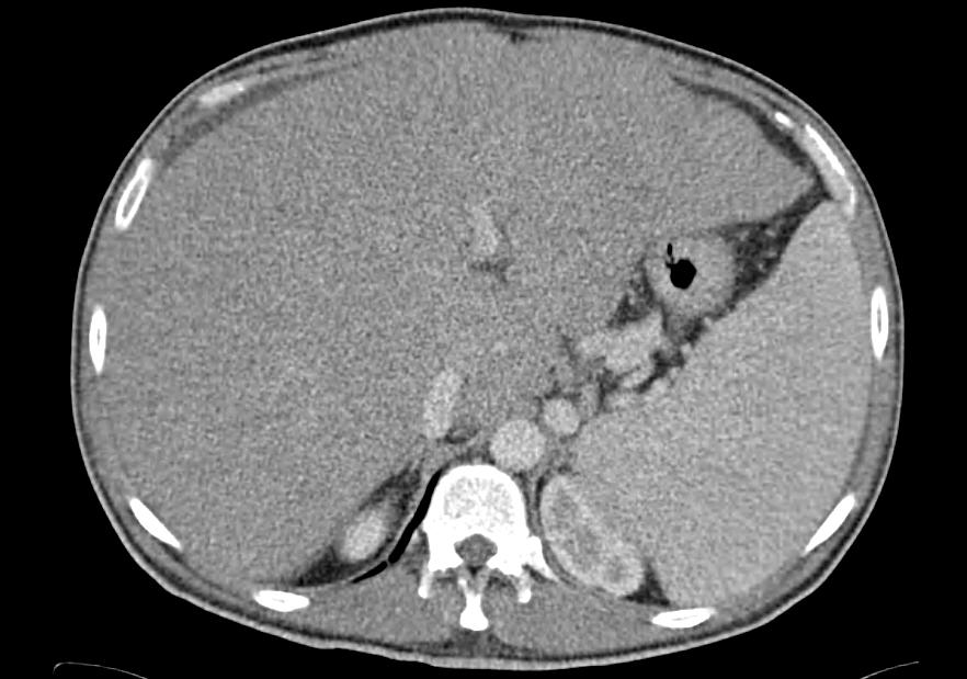
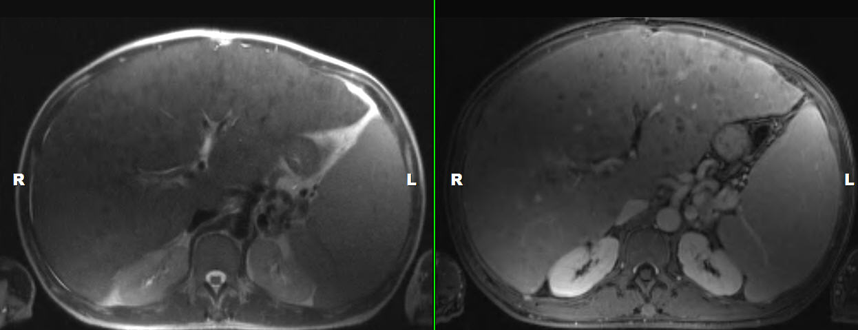
Anatomy: Gastrointestinal (GI)   Pathology: Neoplasm
Modality: MR, Nuc MedAccess Level: Readable by all users, writable by NucMed Certifiers
Keywords: carcinoid

Case has been viewed 33 times.
Certified by Jerold Wallis on 06-24-2009

The reader is fully responsible for confirming the accuracy of this content.
Text and images may be copyrighted by the case author or institution.