Return to Case List with Diagnoses or Case List as Unknowns

LEFT TEMPORAL FIBROUS DYSPLASIA
Authored By: Joanna Fair and Jerold Wallis, Assoc Prof of Radiology.
Patient: 55 year old male
History: Known malignancy (specific history withheld).
Image Size:[small][as-submitted]

Fig. 1
Whole body bone scan.

Fig. 2
Lateral views of the skull on bone scan.

Fig. 3
Oblique rib views on bone scan.

Fig. 4
AP and lateral radiographs of the thoracic spine.

Fig. 5
Temporal bone CT.
Image Size:[small][as-submitted]

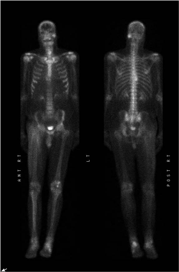
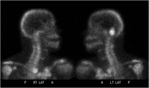
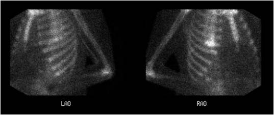
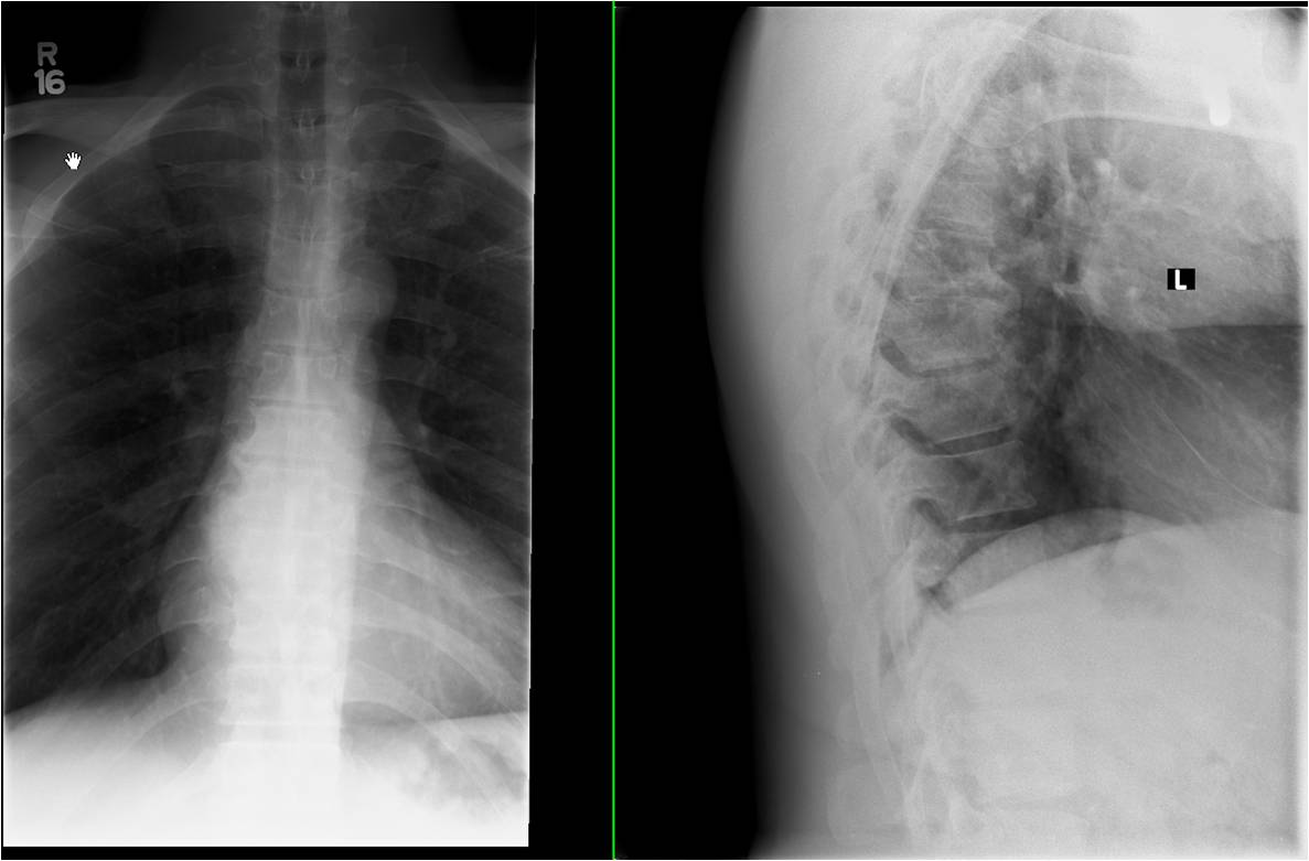
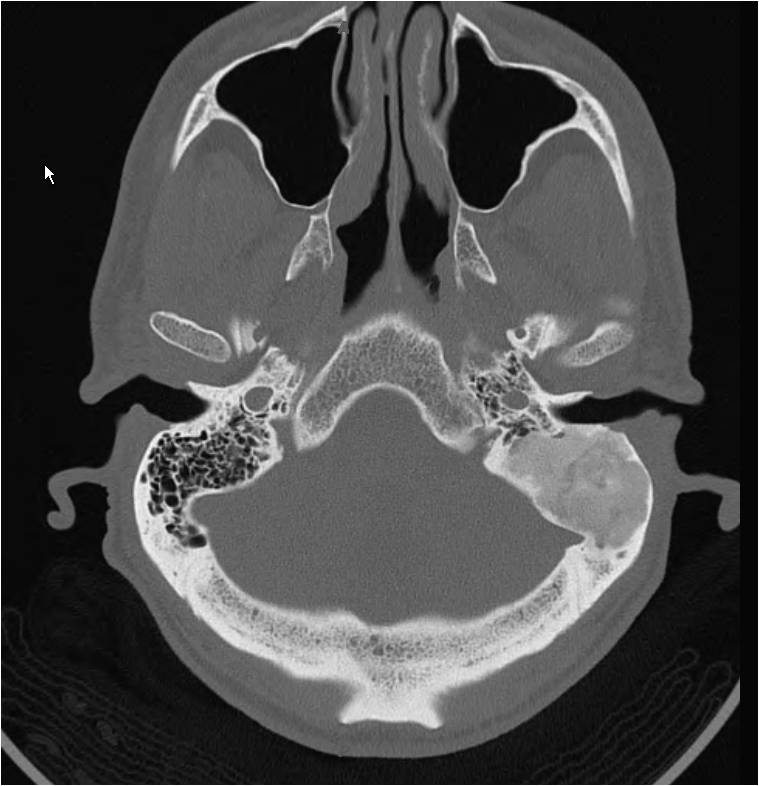
Findings: Radiopharmaceutical:  20.8 mCi Tc-99m MDP i.v.

A whole-body bone scan (Fig. 1) was performed, with additional images of the skull (Fig. 2) and ribs (Fig. 3). 

What are the findings?

[pagebreak]
These images demonstrate focal increased uptake in the region of the left mastoid air cells.  There is moderate diffuse soft tissue uptake in the right anterior proximal thigh, and focal uptake in the mid thoracic spine.

What additional imaging would you obtain?

[pagebreak]

AP and lateral thoracic spine (Fig. 4) radiographs demonstrate degenerative osteophyte formation at the T8-9 level, corresponding to the region of activity on the bone scan.

Temporal bone CT (Fig. 5) demonstrates an expansile lesion of the left petrous temporal bone with ground glass matrix.
DDx: For the left mastoid uptake:  Neoplastic (metastasis versus primary lesion) versus infectious/inflammatory process of the mastoid portion of the left petrous temporal bone vs a benign process such as fibrous dysplasia.
Diagnosis: Fibrous dysplasia of the mastoid portion of the left petrous temporal bone.
General Discussion: Full patient history:

The patient had a right thigh high-grade leiomyosarcoma, diagnosed 1 1/2 years previously, status post surgical resection with adjuvant radiation therapy and chemotherapy.  He was now newly diagnosed with prostate cancer; prostate-specific antigen (PSA) = approximately 15 ng/mL.

The patient also noted a remote history of work-related injury to the mid back.


Discussion:

The CT appearance of the left mastoid lesion is classic for fibrous dysplasia, showing the typical expansile lesion with ground glass matrix.  This location would be unusual for prostate cancer metastasis, particularly given the lack of evidence of metastasis elsewhere.

The soft tissue uptake on the bone scan in the right proximal thigh is consistent with prior surgical resection of and radiation therapy to the soft-tissue sarcoma.

The uptake on the bone scan in the mid thoracic spine is consistent with the degenerative changes seen on the thoracic spine radiographs.
References: Brown EW, Megerian CA, McKenna MJ, Weber A. Fibrous dysplasia of the temporal bone. AJR 1995;164:679-682.

Kransdorf MJ, Moser RP Jr, Gilkey FW. Fibrous dysplasia. RadioGraphics 1990;10:519-537.

Nakahara T, Fujii H, Hashimoto J, Kubo A. Use of bone SPECT in the evaluation of fibrous dysplasia of the skull. Clin Nucl Med. 2004;29(9):554-559.
Comments:
No comments posted.
Additional Details:

Case Number: 130389Owner(s): Joanna Fair and Jerold Wallis, Assoc Prof of RadiologyLast Updated: 02-07-2013
Anatomy: Cranium and Contents   Pathology: Neoplasm
Modality: CT, Conventional Radiograph, Nuc MedAccess Level: Readable by all users, writable by NucMed Certifiers
Keywords: bsnm, fibrous, dysplasia, bone scanACR: 21420.85000

Case has been viewed 47 times.
Certified by Jerold Wallis on 06-24-2009

The reader is fully responsible for confirming the accuracy of this content.
Text and images may be copyrighted by the case author or institution.