Return to Case List with Diagnoses or Case List as Unknowns

EWING'S SARCOMA
Authored By: Farrokh Dehdashti and Xiaoni Hong.
Patient: 12 year old female
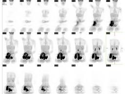
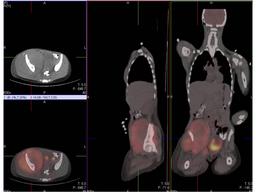
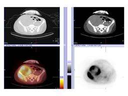
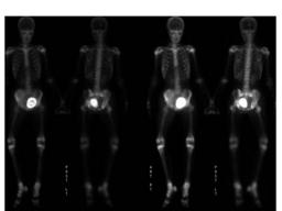
History: 12-year-old female with a right-sided pelvic mass.
Image Size:[small][as-submitted]

Fig. 1
FDG-PET demonstrates markedly increased FDG uptake in the large soft tissue mass in the right lower abdomen and pelvis that is associated with lytic permeative destruction of the right iliac bone. Diffuse bone marrow uptake with more focal increased FDG uptake in several bones involving in the bone marrow throughout the skeleton, with more focal FDG uptake in the L3 and L4 vertebral bodies, left proximal femur, right proximal humerus, as well as left proximal humerus, and right scapula. There is mild focused increased FDG uptake in the right seventh rib anteriorly.

Fig. 2
FDG-PET/CT demonstrates markedly increased FDG uptake in the large soft tissue mass in the right lower abdomen and pelvis that is associated with lytic permeative destruction of the right iliac bone.

Fig. 3
FDG-PET/CT demonstrates markedly increased FDG uptake in the large soft tissue mass in the right lower abdomen and pelvis that is associated with lytic permeative destruction of the right iliac bone.

Fig. 4
Bone scan revearls hazy uptake seen overlying essentially the entire right iliac bone. Decreased uptake in region of the right sacroiliac joint when compared to the left sacroiliac joint. Abnormal uptake in the right acetabular region.Focus of uptake within the right anterolateral 7th rib. A focus of decreased uptake seen within the superior aspect of the left iliac crest without corresponding CT abnormality concerning for possible osseous metastasis.
Image Size:[small][as-submitted]

Findings:

RADIOPHARMACEUTICAL: 6.1 mCi F-18 Fluorodeoxyglucose i.v.
PET/CT deminstrated markedly increased FDG uptake in the large soft tissue mass in the right lower abdomen and pelvis that is associated with lytic permeative destruction of the right iliac bone. Diffuse bone marrow uptake with more focal increased FDG uptake in  the L3 and L4 vertebral bodies, left proximal femur, right proximal humerus, as well as left proximal humerus, and right scapula.


RADIOPHARMACEUTICAL: 11.2 mCi Tc-99m MDP i.v.
Bone scan deminstrated hazy radiotracer uptake overlying essentially the entire right iliac bone. The bladder is displaced to the left. There is asymmetric radiotracer uptake within the right sacroiliac joint (decreased along the superior aspect of the right sacroiliac joint) and the right acetabular region. In addition, there is a focus of radiotracer uptake within the right anterolateral 7th rib with surrounding decreased uptake about this rib on both sides of the increased uptake.

DDx: Ewing's sarcoma
Osteosarcoma
Lymphoma
Rhabdomyosarcoma
Osteomyolitis
Diagnosis: Ewing's sarcoma of the right iliac bone.
General Discussion: Long history: 12-year-old female who presented with right leg pain,
and this developed into a limp which was progressively symptomatic. She had
an associated 20-pound weight loss, more recently developed inability to
urinate. An MRI revealed a 3 cm pelvic mass centered around the right iliac bone.

Discussion: Ewing sarcoma, a highly malignant primary bone tumor, was first described by James Ewing in 1921. The tumor is derived from red bone marrow. Most frequently, it is observed in children and adolescents aged 4-15 years and rarely develops in adults older than 30 years. Ewing sarcoma accounts for approximately 5% of biopsy-analyzed bone tumors and approximately one third of primary bone tumors. Ewing sarcoma is the second most common malignant bone tumor in young patients, and it is the most lethal bone tumor. Males are affected more frequently than females, with a ratio of approximately 1.5:1. Ewing sarcoma occurs in African Americans and Asians. An association exists between Ewing sarcoma and primitive peripheral neuroectodermal tumor. Most frequently, the tumor is diagnosed as a monostotic lesion in the metaphysis or diaphysis of the long bones of the extremities. The tumor also may occur, although less frequently, in the pelvic area, ribs, and scapulae. In fact, any bone may be involved.

Whole-body bone scan can provide information about the primary lesion and depict skip lesions. Also, bone scintigraphy can be used to localize distant metastases during tumor staging.

PET/CT is significantly more accurate than PET alone for the detection and localization of lesions and improves staging for patients with Ewing tumor. The hybrid technique is superior to PET alone in terms of sensitivity, specificity, and accuracy, mainly because of the detection of new lesions. FDG-PET also is useful following therapy, and it can reveal early changes in tumor metabolism, which is an indicator of the therapeutic effect. Bone marrow scanning is of limited use because of the availability of MRI.
References: Völker T, Denecke T, Steffen I, Misch D, Schönberger S, Plotkin M, Ruf J, Furth C, Stöver B, Hautzel H, Henze G, Amthauer H. Positron emission tomography for staging of pediatric sarcoma patients: results of a prospective multicenter trial.
J Clin Oncol. 2007 Dec 1;25(34):5435-41.

Gerth HU, Juergens KU, Dirksen U, Gerss J, Schober O, Franzius C.
 Significant benefit of multimodal imaging: PET/CT compared with PET alone in staging and follow-up of patients with Ewing tumors. J Nucl Med. 2007 Dec;48(12):1932-9.

Hawkins DS, Schuetze SM, Butrynski JE, Rajendran JG, Vernon CB, Conrad EU 3rd, Eary JF.[18F]Fluorodeoxyglucose positron emission tomography predicts outcome for Ewing sarcoma family of tumors. J Clin Oncol. 2005 Dec 1;23(34):8828-34.
Comments:
No comments posted.
Additional Details:

Case Number: 98319Owner(s): Farrokh Dehdashti and Xiaoni HongLast Updated: 02-07-2013
Anatomy: Skeletal System   Pathology: Neoplasm
Access Level: Readable by all users, writable by NucMed Certifiers
Keywords: ptnmACR: 40000.32810

Case has been viewed 51 times.
Certified by Farrokh Dehdashti on 05-22-2008

The reader is fully responsible for confirming the accuracy of this content.
Text and images may be copyrighted by the case author or institution.