Return to Case List with Diagnoses or Case List as Unknowns

HETEROTOPIC OSSIFICATION OF THE RIGHT HIP
Authored By: Keith Fischer and Xiaoni Hong.
Patient: 67 year old female
History:

67-year-old woman with recent cerebral infarction resulting in severe right-sided weakness


Image Size:[small][as-submitted]

Fig. 1
Blood flow images show increased activity around the right hip on the angiographic phase

Fig. 2
Bone scan shows increased activity seen around the right hip on the immediate static, as well as delayed images.

Fig. 3
Plain films show amorphous density seen projecting over the superior aspect of the femoral neck on the AP view and the inferior aspect of the acetabulum on the frog leg view.
Image Size:[small][as-submitted]

Findings:

Bone Scan: There is increased radionuclide seen around the right hip on the angiographic and immediate static, as well as delayed images. This correlates with the region of amorphous calcification seen on the radiograph and is indicative of immature (forming) heterotopic ossification which is very vascular.

Plain film: Amorphous density seen projecting over the superior aspect of the femoral neck on the AP view and the inferior aspect of the acetabulum on the frog leg view.


DDx: Heterotopic ossification
Osteomyolitis
Osteosarcoma
Diagnosis:  heterotopic ossification of the right hip
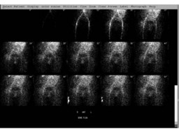
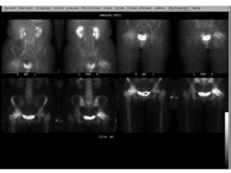
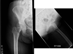
General Discussion:

Long history: 67-year-old woman with recent cerebral infarction resulting in severe right-sided weakness now presenting with amorphous ossification around the right hip on radiographs 06/21/2007. Evaluate for heterotopic ossification.

Radiopharmaceutical: 20.1 mCi Tc-99m MDP i.v.

Discussion: Heterotopic ossification (HO) is the presence of bone in soft tissue where bone normally does not exist. The acquired form of HO most frequently is seen with musculoskeletal trauma, spinal cord injury, or central nervous system injury.  The fever, swelling, erythema, and occasional joint tenderness seen in early HO can be difficult to distinguish from cellulitis, osteomyelitis, or thrombophlebitis. Bone scanning and other imaging tests frequently are used to distinguish between these diagnostic possibilities. Bone scanning may be requested to confirm the diagnosis of HO . In addition, surgical resection of HO is used to preserve joint mobility; however, HO is likely to recur and possibly progress if resection is undertaken before the ossification has become mature. With a view toward avoiding recurrent HO and other operative complications, serial bone scans are used to determine whether medical treatment is warranted, and to choose the appropriate time for surgical resection of HO.


Comments:
No comments posted.
Additional Details:

Case Number: 97104Owner(s): Keith Fischer and Xiaoni HongLast Updated: 02-07-2013
Anatomy: Skeletal System   Pathology: Other
Modality: Nuc MedAccess Level: Readable by all users, writable by NucMed Certifiers
Keywords: bsnmACR: 40000.81500

Case has been viewed 33 times.
Certified by Keith Fischer on 01-29-2009

The reader is fully responsible for confirming the accuracy of this content.
Text and images may be copyrighted by the case author or institution.