back to index of exported cases

Unknown Case
Authored By: Xiaoni Hong and Barry Siegel, Prof of Radiology.
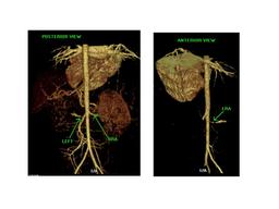
Patient: 2 year 1 month 14 day old male
History: 2-year-old boy admitted with hypertensive emergency.
Image Size:[small][as-submitted]

Fig. 1
Posterior radionuclide angiogram demonstrates decreased perfusion to the smaller left kidney

Fig. 2
Sequential renal scintigrapy images show decreased function of the smaller left kidney, with progressively increasing renal parenchymal uptake of tracer, but no excretion. In light of the clinical history, the findings are most consistent with high-grade left renal artery stenosis. Normal right renal function with no evidence of obstruction.

Fig. 3
Post-voiding image.

Fig. 4
CT angiograhy reveals 6 mm focal stenosis of the left renal artery in its proximal portion. Distal to the stenosis the artery caliber increases to 4 mm in diameter.There is decreased size and a delayed nephrogram of the left kidney.
Image Size:[small][as-submitted]

Comments:
No comments posted.
Additional Details:

Case [View Case with Diagnosis]Owner(s): Xiaoni Hong and Barry Siegel, Prof of RadiologyLast Updated: 02-07-2013
Anatomy: Genitourinary (GU)   Pathology: Vascular
Modality: Nuc MedAccess Level: Readable by all users, writable by NucMed Certifiers
Keywords: rsnmACR: 80000.12170

Case has been viewed 48 times.
Certified by Barry Siegel on 06-13-2009

The reader is fully responsible for confirming the accuracy of this content.
Text and images may be copyrighted by the case author or institution.