Return to Case List with Diagnoses or Case List as Unknowns

PNEUMOTHORAX
Authored By: Brandon Peters and Jerold Wallis, Assoc Prof of Radiology.
Patient: 54 year old male
History: 54 year old man with right sided chest pain.  The patient has a history of multiple sclerosis and has had a recent knee operation and also a colectomy. 
Image Size:[small][as-submitted]

Fig. 1
Xenon 133 lung ventilation images

Fig. 2
Tc-99m MAA lung perfusion images

Fig. 3
Frontal view of the chest

Fig. 4
Frontal view of the chest following chest tube placement
Image Size:[small][as-submitted]

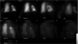
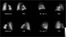
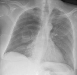
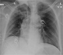
Findings: Xenon 133 lung ventilation images:  There is absent ventilation in a large nonsegment region predominantly in the expected position of a portion of the right upper and middle lobes. Additionally, there is a central photopenic defect medially in the right lung, larger than the expected photopenia attributable to normal hilar structures.  No abnormality of left lung ventilation is seen. 

Tc-99m lung perfusion images:  There is a matching pattern of absent perfusion involving the same nonsegmental region predominantly in the expected position of a portion of the right upper and middle lobes.  There is a matching perfusion defect centrally in the visualized right lung as well.  No abnormalities of the left lung is demonstrated.

Frontal radiograph:  There is a large right pneumothorax with collapse of the right lung medially.

Frontal radiograph following chest tube placement:  Complete resolution of the right pneumothorax with reexpansion of the right lung.
DDx: Matched unilateral nonsegmental, nonlobar ventilation/perfusion defects:

1.  Space occupying lesions:  Pleural effusion, hemothorax, pneumothorax, empyema, pleural neoplasm, plumbage.
Diagnosis: Pneumothorax
Specific Discussion: Although ventilation/pefusion scanning is most often used for evaluating for pulmonary embolus, many other pathologies alter lung ventilation and perfusion which are important to understand when interpreting this type of study.  The hallmark of pulmonary embolus is the segmental mismatched defect (normal ventilation with reduced/absent perfusion).  Matched defects are often thought of as "something that is not pulmonary embolus" but this type of pattern can be further refined.  Pneumonia and other airspace consolidating processes are often lobar or at least multiple contigous segments in a lobe.  Pleural layers serve to limit pneumonia from spreading contigously across lobes.  When a unilateral matched nonsegmental, nonlobar defect is seen, a pleural based process should come to mind.  Although the pleural processes are usually diagnosed by means other than a V/Q study, the symptoms of advanced pleural processes such as shortness of breath, chest pain, and tachycardia are often similar to pulmonary embolus.  Thus, sometimes these processes are discovered on V/Q scans, as in this case.
Comments:
No comments posted.
Additional Details:

Case Number: 94740Owner(s): Brandon Peters and Jerold Wallis, Assoc Prof of RadiologyLast Updated: 02-07-2013
Anatomy: Cardiopulmonary   Pathology: Other
Modality: Conventional Radiograph, Nuc MedAccess Level: Readable by all users, writable by NucMed Certifiers
Keywords: vqnm

Case has been viewed 63 times.
Certified by Jerold Wallis on 10-29-2010

The reader is fully responsible for confirming the accuracy of this content.
Text and images may be copyrighted by the case author or institution.