Return to Case List with Diagnoses or Case List as Unknowns

63-YEAR-OLD WOMAN WITH PAPILLARY THYROID CARCINOMA
Authored By: Farrokh Dehdashti and Asif Moinuddin.
Patient: 63 year old female
History: 63-year-old woman with papillary thyroid carcinoma
Image Size:[small][as-submitted]

Fig. 1

Fig. 2

Fig. 3

Fig. 4

Fig. 5

Fig. 6
Image Size:[small][as-submitted]

Findings:

RADIOPHARMACEUTICAL:  250 mCi therapeutic dose of I-131 sodium iodide

FINDINGS:
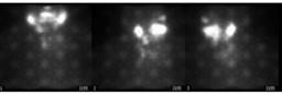
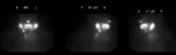
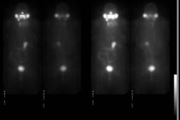
A 250 mCi therapeutic dose of I-131 sodium iodide was administered orally on 04/02/2007 by the staff of the Division of Radiation Oncology. Images of the head, neck, trunk, and proximal extremities were obtained 2 days later.


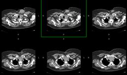
The first set of images obtained were artifactual. Subsequent imaging demonstrated significant portion of the tracer accumulation is within the expected salivary glands, oropharynx and nasopharynx. There are 2 foci of intense tracer accumulations within the right aspect of the thyroid bed consistent with known recurrent disease in this region on prior CT examination. A third larger, less intense focus is seen within the left thyroid bed, which is most likely representative of an area of fluid collection as a result of recent surgical removal of a soft tissue mass from this region. No other lesion is seen. The additional lesions seen on chest CT were not I-131-avid.

Diagnosis: DIAGNOSIS:
Residual functioning thyroid tissue within the thyroid bed, correlating with recurrent thyroid carcinoma

General Discussion: FULL HISTORY:
63-year-old woman with papillary thyroid carcinoma status post thyroidectomy and status post radioactive iodine treatment in 1999, now with finding of recurrence on recent CT examination 02/26/2006 within the thyroid bed, mediastinum and lungs.

DISCUSSION:
The artifact seen on the first image is visualization of the photomultiplier tubes.  This is likely due to inability of the Anger count positioning and correction circuitry to handle high count rate on the gamma camera used for initial imaging. Routinely, I-131 post treatment imaging is performed at 4-5 days post therapeutic dose. This patient was imaged on day 2 and hence, gamma camera was unable to handle the high count rate. Flood fields, photopeaks and images of other studies obtained during that day from the same camera were satisfactory. Later on, patient was imaged on the same day using different gamma camera with thicker crystal and the subsequent images were of diagnostic quality.
Comments:
No comments posted.
Private Notes: ACKNOWLEDGMENT:

Jerry W Wallis, MD
Matthew G Gibson, MD
Additional Details:

Case Number: 94236Owner(s): Farrokh Dehdashti and Asif MoinuddinLast Updated: 02-07-2013
Anatomy: Face and Neck   Pathology: Other
Modality: Nuc MedAccess Level: Readable by all users, writable by NucMed Certifiers
Keywords: trnm, thyroid cancer, variant, artifactACR: 20000.93000

Case has been viewed 44 times.
Certified by Farrokh Dehdashti on 05-22-2008

The reader is fully responsible for confirming the accuracy of this content.
Text and images may be copyrighted by the case author or institution.