|
| ||||||
|
| ||||||
|
| ||||||
|
|
| Patient: 55 year old |
| History: 56 year old male with hypoxia |
Image Size:
|
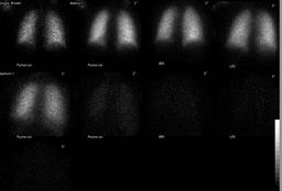
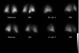
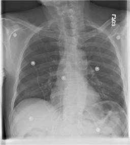
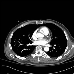
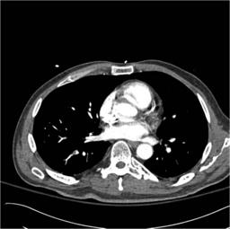
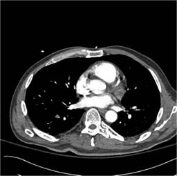
| Findings: Lung ventilation scan:Ā Normal Lung perfusion scan:Ā Moderate sized mismatched defect in the lateral basal segment of the right lower lobe seen on RPO and posterior views.Ā No other defects seen. Chest radiograph:Ā No interstitial or airspace opacities. Chest CT:Ā Thrombus in the lateral segmental artery of the right lower lobe. |
| DDx: Single segmental mismatch: 1.Ā Pulmonary embolus 2.Ā Localized fibrosis |
| Diagnosis: Pulmonary embolus |
| General Discussion: This is a 56 year old male with myasthenia gravis who became acutely hypoxic.ĀThe patient had no previous history of thrombosis or pulmonary embolus. |
| References: Freitas JE.Ā Modified PIOPED criteria used in clinical practice.Ā J Nucl Med 1995;36: 1573-8. |
| Comments: No comments posted. |
| Additional Details:
Case Number: 94186 The reader is fully responsible for confirming the accuracy of this content. |