Return to Case List with Diagnoses or Case List as Unknowns

PTOTIC RIGHT KIDNEY WITH HIGH-GRADE RIGHT URETERAL OBSTRUCTION
Authored By: Xiaoni Hong and Barry Siegel, Prof of Radiology.
Patient: 89 year old female
History: 89-year-old woman with primary peritoneal carcinoma, status post cycle #1 of chemotherapy with carboplatin/Taxol. She was admitted with anemia and dehydration. Her serum creatinine level has increased from 1.1 mg/dL a month ago to 1.8 mg/dL.
Image Size:[small][as-submitted]

Fig. 1
The posterior abdominal radionuclide angiogram demonstrates normal perfusion to the left kidney. The right kidney is ptotic and appears smaller than the left kidney.

Fig. 2
Sequential renal images show the left kidney to be of normal size and morphology. the right kidney appears smaller than the left and is ptotic, again likely related to a large right pleural effusion displacing the right-sided abdominal viscera inferiorly and anteriorly. Activity in the right renal parenchyma increases slowly during the intial 20-minute imaging sequence, and no tracer is seen in the right-sided collecting system.

Fig. 3
The renogram curves show poor function of the right kidney.

Fig. 4
On a post-void image and on anterior and posterior 60-minute delayed images, there is persistent renal parenchymal activity bilaterally. Again, no activity was seen in the right collecting system.

Fig. 5
4.5 hours delayed images demonstrate persistent retention of tracer in the right renal parenchyma and faint persistent parenchymal retention on the left. However, some activity is now seen in the right collecting system and in what appears to be a dilated, tortuous right ureter.

Fig. 6
Axial CT images demonstrate marked, new hydronephrosis of the ptotic right kidney.

Fig. 7
A right-sided was placed after the diagnosis of obstruction was made.
Image Size:[small][as-submitted]

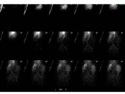
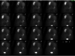
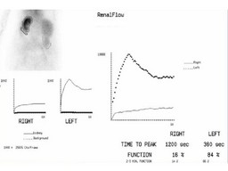
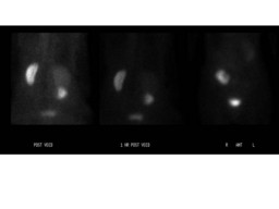
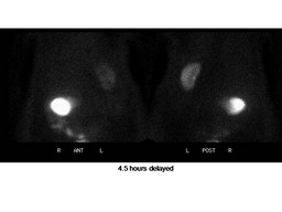
Findings:

Renal Scan: 1. Ptotic right kidney with high-grade right ureteral obstruction, as indicated by markedly delayed excretion and delayed filling of the dilated collecting system and ureter. The relative right renal function is decreased.See general discussion.

2. The parenchymal tracer retention seen on the left raises the question of superimposed medical renal disease and/or prerenal azotemia, as contributing to this patient's new renal insufficiency.


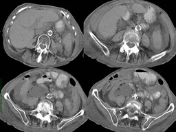
CT findings: Interval development of severe right sided hydronephrosis that appears to terminate at the ureteropelvic junction. This may be due to a metastatic deposit although none is obvious. There is a moderate-sized right-sided pleural effusion and a small left-sided pleural effusion, with a left-sided Pleurex catheter in place.


DDx: Unilateral renal artery stenosis
Unilateral ATN
Unilateral renal vein thrombosis
Unilateral obstruction
Diagnosis: Ptotic right kidney with high-grade right ureteral obstruction
General Discussion: Radiopharmaceutical: 7.9 mCi Tc-99m MAG3 i.v.

Long history: 89-year-old woman with primary peritoneal carcinoma, status post cycle #1 of chemotherapy with carboplatin/Taxol. She was admitted with anemia and dehydration. Her serum creatinine level has increased from 1.1 mg/dL a month ago to 1.8 mg/dL. Evaluate differential renal function and assess for obstruction.

Teaching points: The excretion of the radiopharmacetical into the collecting  system appears markedly delay in this patient who has de hydronephrosis associated with high-grade obstruction. Obtaining delayed image is very important in this situation, to demonstrate delayed filling of the collecting system. Comparison with anatomic imaging studies also is helpful.
Comments:
No comments posted.
Additional Details:

Case Number: 92668Owner(s): Xiaoni Hong and Barry Siegel, Prof of RadiologyLast Updated: 02-07-2013
Anatomy: Genitourinary (GU)   Pathology: Other
Modality: Nuc MedAccess Level: Readable by all users, writable by NucMed Certifiers
Keywords: rsnmACR: 80000.84400

Case has been viewed 30 times.
Certified by Barry Siegel on 06-13-2009

The reader is fully responsible for confirming the accuracy of this content.
Text and images may be copyrighted by the case author or institution.