Return to Case List with Diagnoses or Case List as Unknowns

51-YEAR-OLD MAN WITH LONG-STANDING HISTORY OF SEIZURE ACTIVITY
Authored By: Tom Miller and Asif Moinuddin.
Patient: 51 year old male
History: HISTORY:
51-year-old man with long-standing history of seizure activity.
Image Size:[small][as-submitted]

Fig. 1
FDG-PET Axial images

Fig. 2
FDG-PET coronal images

Fig. 3
FDG-PET Axial images (color maps)

Fig. 4
FDG-PET coronal images (color maps)

Fig. 5
T1-weighted coronal images

Fig. 6
T2-weighted coronal images

Fig. 7
FLAIR coronal images

Fig. 8
FDG-PET and MRI fused image 1

Fig. 9
FDG-PET and MRI fused image 2

Fig. 10
FDG-PET and MRI fused image 3
Image Size:[small][as-submitted]

Findings:

RADIOPHARMACEUTICAL:
10 mCi F-18 Fluorodeoxyglucose i.v.

FINDINGS:
The PET images were fused and compared with the MRI examination dated 8-14-06.There is diffusely decreased FDG uptake within the right temporal lobe, especially pronounced within the right mesial temporal lobe that on MRI images correlates to enlargement in this region along with increased T2 signal abnormality. Note is made of decreased FDG uptake within the cerebellum, which correlates with the patient's use of antiseizure medications.

DDx: DIFFERENTIAL DIAGNOSIS:
Radiation necrosis, recurrent tumor, low- vs. high-grade brain neoplasm
Diagnosis: DIAGNOSIS:
Minimal FDG uptake within the right mesial temporal lobe that on MRI examination correlates to T2 hyperintense right mesial temporal enlargement, most likely representing low-grade tumor.
General Discussion: FULL HISTORY:
51-year-old man with long-standing history of seizure activity. His last seizure activity was in June, 2006. An outside institution MRI examination performed on 7-3-06 was suspicious for infiltrating low-grade temporal lobe neoplasm. PET/CT study is requested for evaluation.

FOLLOW-UP:
The patient underwent right temporal craniotomy for amygdala hippocampectomy and removal of mesial temporal tumor. Pathological examination showed Ganglioglioma; WHO Grade I

Specific Discussion:

DISCUSSION:

WHO classification of Neuroepithelial tumors of the CNS*


1. Astrocytic tumors
2. Oligodendroglial tumors
3. Ependymal cell tumors
4. Mixed gliomas
5. Neuroepithelial tumors of uncertain origin
6. Tumors of the choroid plexus
7. Neuronal and mixed neuronal-glial tumors
 a. Gangliocytoma
 b. Dysplastic gangliocytoma of cerebellum (Lhermitte-Duclos)
 c. GANGLIOGLIOMA
 d. Anaplastic (malignant) ganglioglioma
 e. Desmoplastic infantile ganglioglioma
 f. Desmoplastic infantile astrocytoma
 g. Central neurocytoma
 h. Dysembryoplastic neuroepithelial tumor
 i. Olfactory neuroblastoma (esthesioneuroblastoma)
 j. Variant: olfactory neuroepithelioma
8. Pineal Parenchyma Tumors
9. Tumors with neuroblastic or glioblastic elements (embryonal tumors)

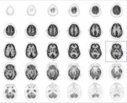
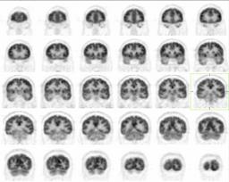
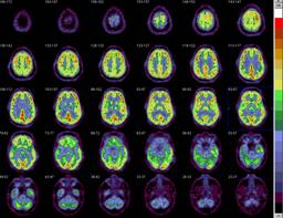
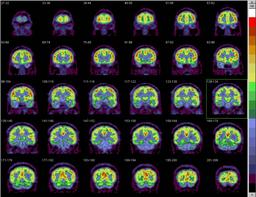
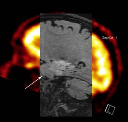
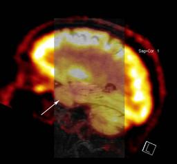
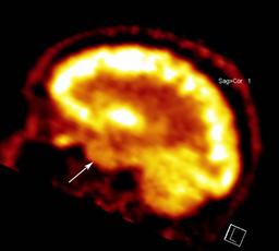
UTILIZATION OF FDG-PET IN TUMOR IMAGING:

FDG-PET in relation to tumor imaging is routinely used for the following pruposes.

1. Differentiating tumor recurrence from post-radiation necrosis
2. Predicting high- vs. low-grade primary brain tumor by evaluating the metobolic activity.
3. Prognostic indicator, by following metabolic activity after treatment. 

References: REFERENCE:
*http://neurosurgery.mgh.harvard.edu/newwhobt.htm
Comments:
No comments posted.
Additional Details:

Case Number: 90706Owner(s): Tom Miller and Asif MoinuddinLast Updated: 02-07-2013
Anatomy: Cranium and Contents   Pathology: Neoplasm
Modality: Nuc MedAccess Level: Readable by all users, writable by NucMed Certifiers
Keywords: ptnm, ganglioglioma, brain, glial tumors, neuronal tumorsACR: 10000.36300

Case has been viewed 64 times.
Certified by Tom Miller on 04-16-2007

The reader is fully responsible for confirming the accuracy of this content.
Text and images may be copyrighted by the case author or institution.