Return to Case List with Diagnoses or Case List as Unknowns

URINARY REFLUX
Authored By: Asif Moinuddin and Jerold Wallis, Assoc Prof of Radiology.
Patient: 1 month 2 day old male
History: 4-week-old boy with urinary tract infection, and recently diagnosed right-sided hydronephrosis.

Image Size:[small][as-submitted]

Fig. 1
Flow

Fig. 2
Split function

Fig. 3
Split graph

Fig. 4
Post-lasix

Multimedia: 85240_5_submitted.avi
Cine during lasix imaging

Fig. 6
Post-lasix graph

Fig. 7
Pre and post lasix
Image Size:[small][as-submitted]

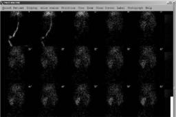
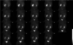
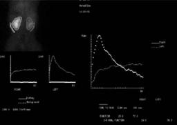
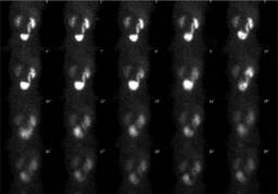
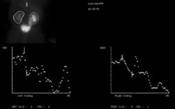
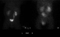
Findings: Normal appearance and function of the left kidney.  Decreased right renal function, as is apparent on the images and the split function curves.

No evidence for obstruction involving the right kidney based on prompt initial post-diuretic clearance of tracer.  However, note the strange shape of the renal curves, with both falling and rising of the curves.

There is a dilated right collecting system and ureter with subsequently observed abrupt increase in activity in the right collecting system, easily cine on the cine images, and also evident on the static display and time-activity curves.  
DDx: Note that patient motion (also present in this case) can cause rising and falling of the renal curves, since patient motion can move the kidney in and out of the drawn region of interest.

In this case, inspection of the cine and static images confirms reflux on the right.

However, rising/falling on the left renal curve appears to be due to patient motion, since motion is present and no left reflux is evident on the cine images.
Diagnosis: Right-sided reflux during the examination. The chronic reflux likely resulted in the mildly diminished right-sided function.
General Discussion: COMPLETE HISTORY:
4-week-old boy with urinary tract infection, and recently diagnosed right-sided hydronephrosis and urinary reflux.

FOLLOW-UP:
The patient was treated with antibiotics.
Specific Discussion: Vesicoureteral reflux (VUR) is defined as retrograde transmission of urine from the urinary bladder to the kidneys The International Reflux Grading system classifies VUR into 5 grades using appearance of the renal pelvis and calyces on VCUG.

Grade 1: Urine backs up into the ureter only, and the renal pelvis appears healthy, with sharp calyces.
Grade 2: Urine backs up into the ureter, renal pelvis, and calyces. The renal pelvis appears healthy and has sharp calyces.
Grade 3: Urine backs up into the ureter and collecting system. The ureter and pelvis appear mildly dilated, and the calyces are mildly blunted.
Grade 4: Urine backs up into the ureter and collecting system. The ureter and pelvis appear moderately dilated, and the calyces are moderately blunted.
Grade 5: Urine backs up into the ureter and collecting system. The pelvis severely dilates, the ureter appears tortuous, and the calyces are severely blunted.

If the goal is specifically to evaluate for reflux using nuclear medicine imaging, this is best done on a nuclear cystogram, with infusion of tracer directly into the bladder after catheterization.  Note that grades 2 and 3 cannot be distinguished on a nuclear medicine study.  

VUR can be a cause of recurrent renal infection in children. VUR can also result in renal failure if left untreated.
Comments:
No comments posted.
Additional Details:

Case Number: 85240Owner(s): Asif Moinuddin and Jerold Wallis, Assoc Prof of RadiologyLast Updated: 02-07-2013
Anatomy: Genitourinary (GU)   Pathology: Congenital
Modality: Nuc MedAccess Level: Readable by all users, writable by NucMed Certifiers
ACR: 80000.85000

Case has been viewed 60 times.
Certified by Jerold Wallis on 05-12-2008

The reader is fully responsible for confirming the accuracy of this content.
Text and images may be copyrighted by the case author or institution.