Return to Case List with Diagnoses or Case List as Unknowns

68-YEAR-OLD-MAN WITH ESOPHAGEAL CANCER
Authored By: Farrokh Dehdashti and Asif Moinuddin.
Patient: 68 year old male
History:

HISTORY:

68-year-old-man with esophageal cancer

Image Size:[small][as-submitted]

Multimedia: 85219_1_submitted.avi

Fig. 2

Fig. 3
Image Size:[small][as-submitted]

Findings:

FINDINGS:

There is a 2 cm right upper lobe, mildly FDG-avid mass, with a maximum standardized uptake value (SUV) of 3.6. This may represent a primary bronchogenic cancer or metastatic esophageal cancer.

DDx:

DIFFERENTIAL DIAGNOSIS:

Lung cancer, metatstatic cancer, esophageal cancer, infection, inflammation

Diagnosis: DIAGNOSIS:
PULMONARY ABSCESS
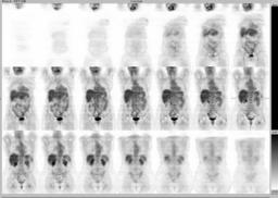
General Discussion:

RADIOPHARMACEUTICAL: 14.6 mCi F-18 Fluorodeoxyglucose i.v.

FULL HISTORY:

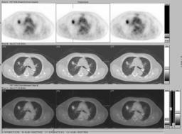
68 year old man with esophageal cancer, status post gastric pull-through procedure, June 2002. There was one metastatic lymph node at the time of surgery. He recently had a new, 2 cm right upper lobe mass diagnosed on follow-up CT. This has developed within 6 months since the last chest CT. Evaluate with PET/CT for restaging esophageal cancer.


FOLLOW-UP:

The patient underwent right upper lobectomy. The lesion was a well circumscribed mass measuring 4.0 cm in greatest dimension, with the bulk represented by interstitial fibrosis with foci of bronchial obliteration. The cystic areas contain necroinflammatory debris with no features of malignancy. No fungal organisms are seen. Final diagnosis, pulmonary abscess with acute and chronic inflammation and reactive interstitial fibrosis.

Follow-up chest CT in 7 months (May 2005) was negative for recurrent or metastatic disease.
Specific Discussion: Teaching Point:
Common false positive FDG-avid lesions include infection and inflammation.
References: Chang JM, Lee HJ, Goo JM, Lee HY, Lee JJ, Chung JK, Im JG.  False positive and false negative FDG-PET scans in various thoracic diseases. Korean J Radiol. 2006 Jan-Mar;7(1):57-69. Review.

Bryant AS, Cerfolio RJ.  The maximum standardized uptake values on integrated FDG-PET/CT is useful in differentiating benign from malignant pulmonary nodules. Ann Thorac Surg. 2006 Sep;82(3):1016-20.
Comments:
No comments posted.
Additional Details:

Case Number: 85219Owner(s): Farrokh Dehdashti and Asif MoinuddinLast Updated: 02-07-2013
Anatomy: Cardiopulmonary   Pathology: Infection
Modality: Nuc MedAccess Level: Readable by all users, writable by NucMed Certifiers
Keywords: ptnm, abscess, pulmonary, noduleACR: 60000.21000

Case has been viewed 64 times.
Certified by Farrokh Dehdashti on 05-22-2008

The reader is fully responsible for confirming the accuracy of this content.
Text and images may be copyrighted by the case author or institution.