Return to Case List with Diagnoses or Case List as Unknowns

38-YEAR-OLD WOMAN WITH BREAST CANCER
Authored By: Keith Fischer and Asif Moinuddin.
Patient: 38 year old female
History: HISTORY:  38-year-old woman, status post right mastectomy for invasive ductal breast cancer.
Image Size:[small][as-submitted]

Fig. 1

Fig. 2

Fig. 3

Fig. 4

Fig. 5
Image Size:[small][as-submitted]

Findings: RADIOPHARMACEUTICAL: 21.4 mCi Tc-99m MDP i.v.

FINDINGS:
Focal area of increased uptake in the first sternal segment Figs 1 and 2), which correlates to a mixed lytic/blastic lesion seen on a contemporary CT examination (Figs 3-5). Bilateral clavicular heads at the sternoclavicular joints also show focally increased uptake, right greater than the left. This may indicate bilateral underlying degenerative changes but a metastasis on the right cannot be excluded. A small focal area of increased uptake within the anterior, inferior end plate of L4, which may represent a metastatic focus or degenerative change is also seen.
DDx: Osseous metastatic disease, post-radiation change, degenerative disease
Diagnosis: RADIATION OSTEITIS
General Discussion:

FULL HISTORY:

38-year-old woman, status post right mastectomy for invasive ductal breast cancer. In addition, she underwent radiation for supraclavicular nodal recurrence. She now presents with sternal and chest wall pain. A recent chest CT scan demonstrated a mixed lytic/sclerotic lesion involving the right clavicular head. Evaluate for osseous metastatic disease. 


FOLLOW-UP:
FDG-PET performed the same day was negative for osseous metastatic disease or other evidence of metastases. Subsequent pathologic examination showed marrow fibrosis, with scattered mast cells, without any significant acute or chronic inflammatory infiltrate and no evidence of metastatic carcinoma. Overall, these findings were most consistent with so-called radiation osteitis. 3-month follow-up bone scintigraphic examination was stable (images not shown)

Specific Discussion: Radiation osteitis was first described by Ewing in 1926. Radiation causes inflammation and necrosis of the marrow space. Radiographically, these changes become evident after 1 to 2 years post radiation, and are manifested by minimal osteoporosis in the radiation treatment zone. Later, trabecular coarsening, patchy sclerosis, and focal areas of destruction occur. The minimal radiation dose to cause this phenomenon is 1500 rad, with a mean dose in the range of 4000 to 5000 rad usually being responsible.
References: Essentials of Radiology: Paul and Juhl's; 7th edition, pages 265-267
Comments:
No comments posted.
Additional Details:

Case Number: 85192Owner(s): Keith Fischer and Asif MoinuddinLast Updated: 02-07-2013
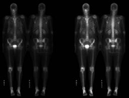
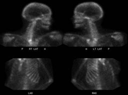
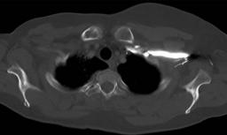
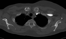
Anatomy: Skeletal System   Pathology: Non-Infectious Inflammatory Disease
Modality: Nuc MedAccess Level: Readable by all users, writable by NucMed Certifiers
Keywords: bsnm, radiation, osteitis, inflammation, metastates, paget'sACR: 40000.47000

Case has been viewed 41 times.
Certified by Keith Fischer on 01-27-2009

The reader is fully responsible for confirming the accuracy of this content.
Text and images may be copyrighted by the case author or institution.