Return to Case List with Diagnoses or Case List as Unknowns

CROSS CEREBELLAR DIASCHISIS
Authored By: Farrokh Dehdashti and Asif Moinuddin.
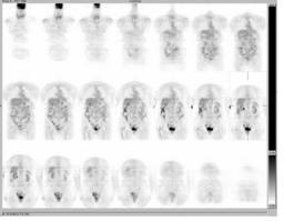
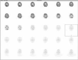
Patient: 68 year old female
History: 68-year-old woman with history of metastatic breast carcinoma to the bones.
Image Size:[small][as-submitted]

Fig. 1
No evidence of recurrent disease.

Fig. 2
Limited examination of the base of the brain demonstrates asymmetric uptake FDG with slightly decreased activity within the left occipital lobe. In addition, there is asymmetric decreased activity within the right cerebellum, compatible with crossed cerebellar diaschisis.

Fig. 3
Limited examination of the base of the brain demonstrates asymmetric decreased activity within the right cerebellum, compatible with crossed cerebellar diaschisis.

Fig. 4
Limited examination of the base of the brain demonstrates asymmetric uptake FDG with slightly decreased activity within the left occipital lobe. There is also evidence of volume loss with ex-vacuole dilatation of the occipital horns of the lateral ventricles, greater on the left. This is compatible with history of old left occipital lobe infarct.
Image Size:[small][as-submitted]

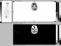
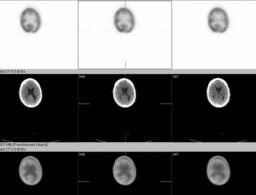
Findings: Limited examination of the base of the brain demonstrates asymmetric FDG uptake, with slightly decreased activity within the left occipital lobe. In addition, there is evidence of volume loss with ex-vacuole dilatation of the occipital horns of the lateral ventricles, greater on the left. This is compatible with history of old left occipital lobe infarct. There also is asymmetric decreased activity within the right cerebellum, compatible with crossed cerebellar diaschisis.
Diagnosis: CROSS CEREBELLAR DIASCHISIS
General Discussion:

Crossed cerebellar diaschisis (CCD) is hypometabolism of cerebellar hemisphere, that is contralateral to the supratentorial lesion resulting in decreased in metabolism and/or perfusion. This mostly occurs as a result of infarction involving the corticopontocerebellar connections or tumor involvement. Frequently, CCD is seen in patients with infarctions of the frontoparietal lobe, basal ganglia, and internal capsule. This phenamenon can be seen in metabolism/perfusion PET or SPECT imaging of the brain.


Specific Discussion:

Acknowledgement:
LIU, JUDY SHENG-SEN
 

References: Kim SE et al. Crossed-cerebellar diaschisis in cerebral infarction: JNM 1997 Dec; 38(12):14-19
Kushner M et al. Contralateral cerebellar hypometabolism following cerebral Infarction: Neurology, 1988;38:147
Comments:
No comments posted.
Additional Details:

Case Number: 85173Owner(s): Farrokh Dehdashti and Asif MoinuddinLast Updated: 02-07-2013
Anatomy: Cranium and Contents   Pathology: Vascular
Modality: Nuc MedAccess Level: Readable by all users, writable by NucMed Certifiers
Keywords: fdg, brain, diaschisis, strokeACR: 10000.70000

Case has been viewed 45 times.
Certified by Farrokh Dehdashti on 07-07-2008

The reader is fully responsible for confirming the accuracy of this content.
Text and images may be copyrighted by the case author or institution.