Return to Case List with Diagnoses or Case List as Unknowns

BILE LEAK
Authored By: Xiaoni Hong and Jerold Wallis, Assoc Prof of Radiology.
Patient: 64 year old male
History: 64 year old man with prior liver transplant, now with abdominal pain.
Image Size:[small][as-submitted]

Fig. 1
First hour HIDA images show progressively increasing activity in the gallbladder fossa.

Fig. 2
90 minute delayed images: Increasing activity located in the gallbladder fossa.

Fig. 3
transaxial CT images show fluid collection along the undersurface of the right lobe of the liver ansd ascites.

Multimedia: 83527_4_submitted.avi
Cine image of the HIDA shows progressively increasing activity over time in the gallbladder fossa.
Image Size:[small][as-submitted]

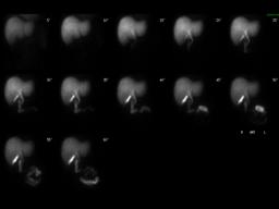
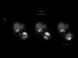
Findings:

HIDA: There is prompt, uniform accumulation of the tracer by the liver. There is normal filling of the intrahepatic ducts, common bile duct and normal excretion of the tracer into the duodenum. Intense tracer accumulation is seen in the gallbladder fossa.

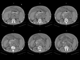
CT: Heterogeneous collection along the undersurface of the right lobe of the liver, likely acute or older hematoma. Small amount of intraperitoneal hemorrhage in right paracolic gutter down to pelvis. Large amount of ascites.

Ultrasound: Ascites. Fluid collection at the inferior aspect of the right hepatic lobe (image not show here).

DDx: 1.Normal gallbladder
2.Loculated bile leak in the gallbladder fossa in a patient status post cholecystectomy
Diagnosis: Loculated bile leak, simulating appearance of gallbladder.
General Discussion: Long History:

64 s/p orthotopic liver transplant for end stage liver disease, due to primary biliary cirrhosis with portal vein thrombosis.  The gallbladder was removed as part of the transplant procedure.   Immediate post-op course was uneventful with appropriate decrease in INR and LFTs. Discharged POD#4

3 weeks after transplant presented with diffuse abdominal pain.
Labs: WBC 20.8, T-Bili 1.6, Alk Phos 192, AST 23, ALT 40

Radiopharmaceutical: 3.1 mCi Tc-99m mebrofenin i.v.

Increasing activity in the gallbladder fossa in this patient who is status post cholecystectomy is consistent with a loculated bile leak.
Specific Discussion: Follow up:
The patient underwent operative procedure which demonstrated several liters of dark colored ascites, with a loculated fluid collection approximately 50 to 100 ml and bile staining in the right subhepatic space. Perforation of the bile duct in the region of the donor cystic duct was seen at surgery, and was reconstructed with a Roux en Y hepaticojejunostomy. The liver parenchyma was normal in appearance.
Comments:
No comments posted.
Additional Details:

Case Number: 83527Owner(s): Xiaoni Hong and Jerold Wallis, Assoc Prof of RadiologyLast Updated: 02-07-2013
Anatomy: Gastrointestinal (GI)   Pathology: Other
Modality: CT, Nuc MedAccess Level: Readable by all users, writable by NucMed Certifiers
Keywords: hpnmACR: 70000.28000

Case has been viewed 66 times.
Certified by Jerold Wallis on 04-05-2007

The reader is fully responsible for confirming the accuracy of this content.
Text and images may be copyrighted by the case author or institution.