Return to Case List with Diagnoses or Case List as Unknowns

REVERSE VENTILATION-PERFUSION MISMATCHING
Authored By: Xiaoni Hong and Barry Siegel, Prof of Radiology.
Patient: 15 year old female
History: 35-year-old woman with sepsis and hypoxia.  The patient is on a ventilator.  Evaluate for pulmonary embolism.
Image Size:[small][as-submitted]

Fig. 1
This is a portable study performed in the intensive care unit. The upper row is aerosol ventilation imaging. The lower two rows are perfusion imaging. The Tc-99m DTPA aerosol images show absence of ventilation to the entire left lung, except for a small region centrally. There is well-maintained perfusion to the entire left lung.

Fig. 2
Portable chest radiographs show misplacement of endotracheal tube into the right main bronchus at 1:40. There is significant left-sided atelectasis.
Image Size:[small][as-submitted]

Findings:

Ventilation-perfusion scintigraphy: The Tc-99m DTPA aerosol images show absence of ventilation to the entire left lung, except for a small region centrally. There is hyperdeposition of the aerosol in the intubated trachea and in the left main bronchus. The perfusion images show a physiologic distribution of pulmonary perfusion. Specifically, perfusion is well maintained to the non-ventilated left lung.
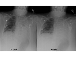
The chest radiograph initially showed significant left sided atelectasis. After the patient was intubated, a repeat radiograph showed good position of the endotracheal tube and persistence of the left-sided volume loss. The patient had not improved and the lung scintigraphy was ordered.

Reverse ventilation-perfusion mismatching (absent perfusion with maintained perfusion) in the entire left lung. The findings indicate substantial functional right-to-left shunting and likely explain the patient's hypoxia.


DDx: DDX: Misplacement of endotracheal tube
        Mucous plug
        Endobronchial lesion
        Extrinsic bronchial compression
        Atelectasis
        Pleural effusion
Diagnosis: Reverse ventilation-perfusion mismatching (absent perfusion with maintained perfusion).  There is failure of expected hypoxic vasoconstriction in the nonventilated left lower lobe.
General Discussion: RADIOPHARMACEUTICAL: Less than 2 mCi Tc-99m DTPA aerosol by inhalation and 5.1 mCi Tc-99 MAA i.v.

Long history: 35-year-old female who was admitted with pneumonia and acute respiratory distress.  She is on a ventilator. Evaluate for pulmonary embolism.

Discussion: Reverse ventilation-perfusion mismatching indicates physiologic right to left shunting. Blood passes through the pulmonary capillary bed without being oxygenated. This is frequently seen in intubated, intensive care unit patients, often the result of central mucous plugging. Those can cause significant hypoxia.

This patient's problem was caused by misplacement of endotracheal tube into the right main bronchus. Certain measures can be taken in this situation. Putting the patient in the decubitus position with the mismatched side up (in this case, left side up) will gravitationally decrease blood flow to affected lung. If PEEP is being used to help ventilate the patient, this can exacerbate the problem by relatively decreasing flow to the ventilated lung, while the poorly ventilated area receives a greater portion of the total pulmonary flow (since the PEEP pressures are transmitted slightly less efficiently to the obstructed region). If possible, positive end-expiratory pressure should be turned off in patients with evidence of functional right-to-left shunting. Finally, the primary problem should be directly addressed, e.g., reposition the endotracheal tube, remove mucous plug by suctioning or bronchoscopy.

Comments:
No comments posted.
Additional Details:

Case Number: 81037Owner(s): Xiaoni Hong and Barry Siegel, Prof of RadiologyLast Updated: 02-07-2013
Anatomy: Other   Pathology: Other
Modality: Nuc MedAccess Level: Readable by all users, writable by NucMed Certifiers
Keywords: vqnmACR: 60000.12176

Case has been viewed 60 times.
Certified by Barry Siegel on 06-09-2009

The reader is fully responsible for confirming the accuracy of this content.
Text and images may be copyrighted by the case author or institution.