Return to Case List with Diagnoses or Case List as Unknowns

LINGUAL THYROID
Authored By: Asif Moinuddin and Jerold Wallis, Assoc Prof of Radiology.
Patient: 26 year old female
History:

HISTORY:

26-year-old woman with hypothyroidism


Image Size:[small][as-submitted]

Fig. 1
Thyroid Scintigraphy (Anterior, with pinhole collimator at 4 cm from neck)

Fig. 2
Thyroid Scintigraphy (Anterior, with pinhole collimator at 14 cm from neck). The right-most image has markers denoting the level on the patient.

Fig. 3
Thyroid Scintigraphy (Lateral; emission and transmission)
Image Size:[small][as-submitted]

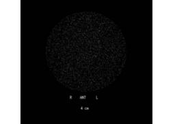
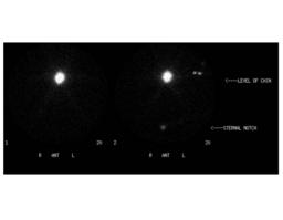
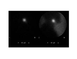
Findings: RADIOPHARMACEUTICAL: 0.25 mCi I-123 sodium iodide capsule

FINDINGS: Pinhole images of the anterior neck (fig. 1) do not demonstrate any thyroid tissue in the expected location of the thyroid. Images obtained anteriorly at a greater distance from the patient (fig. 2) demonstrate a focus of activity just inferior to the chin.  Additional lateral images (fig. 3) using a parallel hole collimator show the uptake to be located at the base of the tongue.  The transmission images were obtained by holding a Co-57 sheet source behind the patient during imaging, which helps significantly in anatomic localization of the thyroid tissue.
Diagnosis: Ectopic Thyroid Tissue (lingual thyroid)  In this case, it is the only thyroid tissue the patient has.
General Discussion:

DISCUSSION:

Several ectopic locations for thyroid tissue have been described.  Ectopic thyroid tissue can occur anywhere from the base of the tongue to the substernal region. Ectopic tissue has been discribed in tumors of the pelvis (struma ovarii).  Tc-99m pertechnetate imaging would be less useful in detecting these ectopic locations because of salivary gland uptake, soft tissue or bony attenuation and blood pool activity. Radioactive iodine (I-123) imaging at 24 hours post administration allows more time for clearance of soft tissue activity and is more helpful when exploring these deep ectopic locations for suspected ectopic thyroid tissue.  Use of I-123 minimizes radiation dose, and is preferred over I-131 in this clinical setting because it has no beta emissions.


References: *Fred A Mettler, Jr. and Milton J. Guiberteau; Essentials of Nuclear Medicine Imaging, 5th edition, 2006
Comments:
No comments posted.
Additional Details:

Case Number: 79019Owner(s): Asif Moinuddin and Jerold Wallis, Assoc Prof of RadiologyLast Updated: 02-07-2013
Anatomy: Face and Neck   Pathology: Normal/Variants
Modality: Nuc MedAccess Level: Readable by all users, writable by NucMed Certifiers
Keywords: trnm, lingualACR: 20000.14750

Case has been viewed 122 times.
Certified by Jerold Wallis on 05-12-2008

The reader is fully responsible for confirming the accuracy of this content.
Text and images may be copyrighted by the case author or institution.