Return to Case List with Diagnoses or Case List as Unknowns

NONOBSTRUCTING/NONREFLUXING PRIMARY MEGAURETER
Authored By: Keith Fischer and Rosana Ponisio.
Patient: 1 month 13 day old
History: 44-day-old baby boy with right hydronephrosis and hydroureter.
Image Size:[small][as-submitted]

Fig. 1
Figure 1: posterior abdominal radionuclide angiogram

Fig. 2
Figure 2: posterior images (20 minutes)

Fig. 3
Figure 3:To evaluate for obstruction, the patient was given furosemide.

Fig. 4
Figure 4:Time activity curve

Fig. 5
figure 5: Renal sonogram ( right kidney)

Fig. 6
Figure 6: voiding cystourethrogram
Image Size:[small][as-submitted]

Findings:

Diuretic renal scintigraphy was performed (1.2 mCi Tc-99m MAG3 i.v. and 5 mg furosemide i.v.).

The posterior abdominal radionuclide angiogram demonstrates normal, symmetrical perfusion of the kidneys (figure 1). After 20 minutes of imaging, there is a small amount of retained activity in the left collecting system, which appears normal in size. There is a moderate amount of retained activity in the right collecting system, which appears moderately enlarged. There is faint tracer activity in the dilated right renal pelvis and proximal ureter. The left ureter appears normal. The right ureter appears tortuous and dilated. The bladder appears normal (figure 2).

To evaluate for obstruction, the patient was given furosemide. Sequential images were obtained for an additional 20 minutes with the patient in the supine position. There is prompt clearance of pelvicalyceal activity on the left after diuretic administration. On the right, there is also prompt clearance of activity from the pelvicalyceal system (figure 3). After diuretic administration, the half-time of tracer clearance from the left kidney was not calculated since there was no residual tracer activity in the renal collecting system. The half-time of tracer clearance from the right kidney is 7 minutes and from the right dilated ureter is 7 minutes (figure 4).

The patient had a renal sonogram that demonstrated moderate to severe right hydronephrosis and hydroureter (figure 5), and a voiding cystourethrography study was normal without evidence of vesicoureteral reflux (figure 6).

DDx:

Differential Diagnosis:

1. Congenital Megaureter

2. Vesicoureteral reflux

3. Duplex collecting system.

Diagnosis:

Diagnosis:

Non-refluxing unobstructed primary megaureter

General Discussion:


 The term congenital (primary) megaureter includes causes of an enlarged ureter which are intrinsic to the ureter, rather than as a result of a more distal abnormality. A ureter larger than 5 mm in diameter should be considered abnormal in childhood.  The megaureters can be obstructing, refluxing or refluxing/nonobstructing.

 The obstructive megaureter is related to a distal adynamic segment with proximal dilatation, and is a common cause of obstructive uropathy in children. It is analogous to esophageal achalasia or colonic Hirschsprung disease although lack of ganglion cells within the wall of the ureter has not been proven to be the cause.

Refluxing primary megaureter is a result of an abnormal vesico-ureteric junction, which impedes the normal anti-reflux mechanisms. This can be due to a short vertical intramural segment, congenital paraureteric diverticulum, ureterocoele with or without associated duplicated collecting system

Non-refluxing unobstructed primary megaureter is thought to be the most common cause of primary megaureter in neonates, and even though the vesicoureteric junction is normal, with no evidence of reflux or obstruction, the ureter is enlarged. The cause for this is unknown.

Bilateral involvement is present in about 20% of patients with primary obstructed megaureters. Primary obstructed megaureter has a male-to-female ratio of nearly 4:1. The left side is more often affected than the right.

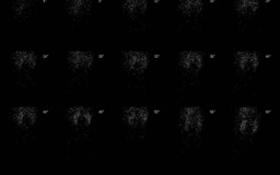
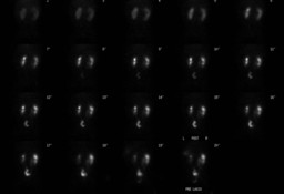
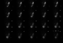
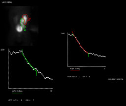
Technetium-99m mercaptoacetyltriglycine (MAG3) is the most widely employed radiopharmaceutical due to the low neonatal GFR. The diuretic renal scintigraphy is a useful test in determining whether a dilated collection system is functionally obstructed. The evaluation of different parameters, including differential renal uptake and time to half peak after furosemide washout, allows a reliable assessment of obstruction to the urinary flow.

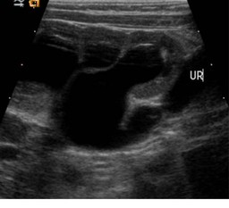
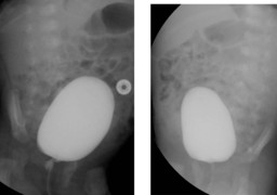
Imaging studies such as ultrasound, voiding cystourethrography and renal scan can help to differentiate primary megaureter from other causes of hydronephrosis, including ureteropelvic junction obstruction, posterior urethral valves, and ureterocele.

The treatment for asymptomatic patients with non-refluxing unobstructed primary megaureter and normal renal scan, as in the present case is nonoperative management because, in most cases, hydrouteronephrosis resolves spontaneously


References:

 The Diagnosis and Treatment for Primary Obstructive Megaureter in Adult and Children-Three Cases Experience and Review of the Literatures; Siu-San Tse, Jue-Hawn Yin; JTUA (2009) 20:173-7.

 Education article: Primary non-refluxing megaureters; E. Merlini, P. Spina; "Journal of Pediatric Urology" (2005) 1, 409-417.

http://www.uptodate.com/contents/primary-megaureter-in-infants-and-children

Comments:
No comments posted.
Additional Details:

Case Number: 446205Owner(s): Keith Fischer and Rosana PonisioLast Updated: 02-07-2013
Anatomy: Genitourinary (GU)   Pathology: Congenital
Modality: Nuc Med, GU, USAccess Level: Readable by all users, writable by NucMed Certifiers
Keywords: primary megaureter, renal scan, rsnm

Case has been viewed 15 times.
Certified by Keith Fischer on 02-04-2013

The reader is fully responsible for confirming the accuracy of this content.
Text and images may be copyrighted by the case author or institution.