Return to Case List with Diagnoses or Case List as Unknowns

URETEROPELVIC JUNCTION OBSTRUCTION WITH MASSIVE DILATATION OF THE RENAL COLLECTING SYSTEM
Authored By: Keith Fischer and Rosana Ponisio.
Patient: 15 year old female
History: Previously healthy, 15 year old  girl who presented with a 2-day history of left abdominal pain, vomiting, headache, and fever.
Image Size:[small][as-submitted]

Fig. 1
Figure 1:posterior abdominal radionuclide angiogram

Fig. 2
Figure 2: Sequential renal images the kidneys (20 minutes)

Fig. 3
Figure 3

Fig. 4
Figure 4:

Fig. 5
Figure 5: half-time of tracer clearance after diuretic administration

Fig. 6
Figure 6 : renal sonogram (left kidney)

Fig. 7
Figure 7: CT with intravenosus contrast ( coronal view)
Image Size:[small][as-submitted]

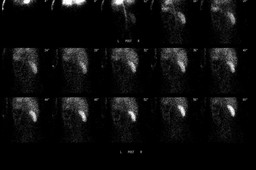
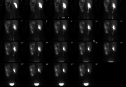
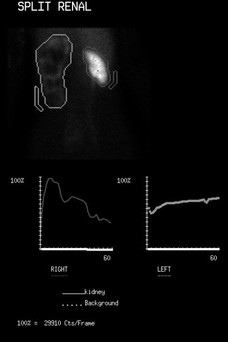
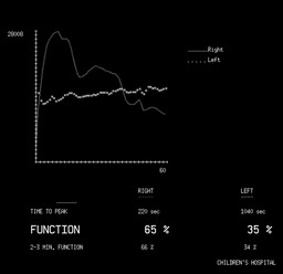
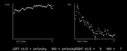
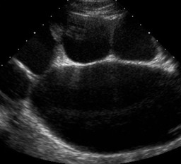
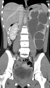
Findings:

Diuretic renal scintigraphy: The posterior abdominal radionuclide angiogram demonstrates decreased perfusion of the left kidney and normal  right renal perfusion(Fig 1). Sequential renal images:  The left kidney and its extrarenal pelvis are massively enlarged and and the parenchyma is thin with prompt uptake and excretion of the radiopharmaceutical by both kidneys (Fig 2).

The estimated contribution of the right kidney to total renal function is 65% and that of the left kidney is 35% (Fig 3, 4). There is no appreciable clearance of pelvicalyceal activity on the left after diuretic administration. On the right, there is normal, rapid clearance of activity from the pelvicalyceal system.

 After diuretic administration, the half-time of tracer clearance from the right kidney is 5 minutes and from the left kidney is unmeasurable due to lack of clearance (Fig  5).

 The renal sonogram as well as the computed tomography demonstrate left ureteropelvic junction obstruction with marked dilation of the left renal pelvis and calyces (Figure 6, 7)

DDx:

Differential diagnosis:

1.  Left ureteropelvic junction obstruction and massive dilatation of the renal collecting system

2.   Hydronephrosis without obstruction

Diagnosis:


1.  Left ureteropelvic junction obstruction and massive dilatation of the renal collecting system

General Discussion:

Discussion:

Ureteropelvic junction (UPJ) obstruction is defined as an obstruction of the flow of urine from the renal pelvis to the proximal ureter. The condition is encountered in both adult and pediatric population. In adults, other etiologies for ureteral obstruction must be considered, including stones, ureteral compression due to extrinsic processes, retroperitoneal fibrosis, and other inflammatory processes and can present with various symptoms, including back and flank pain, UTI, and/or pyelonephritis.

In cases of hydronephrosis caused by obstruction at the ureteropelvic junction or ureterovesical junction, dynamic renal scintigraphy demonstrates abnormalities in structure and function on the involved side. On dynamic studies, the hydronephrotic kidney initially appears as a rim of tracer concentration in the renal parenchyma surrounding an area devoid of tracer, corresponding to the renal pelvis and collecting system. The rate of appearance and the amount of the tracer in the pelvicaliceal system depends on the function of the hydronephrotic kidney

The apparent function of the hydronephrotic kidney depends on the degree of urinary obstruction that increases the pressure in the pelvicaliceal system and the presence and degree of renal damage.  The level of obstruction is inferred by the renal and collecting system washout curves. Low grade of partial obstruction demonstrates a half-time of tracer clearance of 10-20 minutes.  In high grade  obstruction, the half-time of tracer clearance is greater than 20 minutes.   False positive or indeterminate diuretic washout times may be seen in poor renal function, poor response to lasix, severely dilated but non-obstructed collecting systems, immature kidneys, or dehydration.


References:

Rossleigh MA et al: Determination of the normal range of furosemide half-clearance times when using Tc-99m MAG3. Clin Nucl Med. 19(10):880-2, 1994.

 Karam M et al: Diuretic renogram clearance half-times in the diagnosis of obstructive uropathy: effect of age and previous surgery. Nucl Med Commun. 24(7):797-807, 2003

 "Pediatric Nuclear Medicine/PET" : Salvador T. Treves; Springer (2007) third edition


Comments:
No comments posted.
Additional Details:

Case Number: 373641Owner(s): Keith Fischer and Rosana PonisioLast Updated: 02-07-2013
Anatomy: Genitourinary (GU)   Pathology: Congenital
Modality: Nuc Med, USAccess Level: Readable by all users, writable by NucMed Certifiers
Keywords: ureteropelvic junction obstruction,hydronephrosis, rsnm

Case has been viewed 16 times.
Certified by Keith Fischer on 01-28-2013

The reader is fully responsible for confirming the accuracy of this content.
Text and images may be copyrighted by the case author or institution.