|
| Patient: 8 year old male |
| History: 8-year-old boy with no significant past medical history who presents after being struck in the head by a dodge ball approximately 2 months ago.
On physical exam, there is a 2 x 2 cm area of swelling around the left mastoid in the post-auricular region with no active drainage from the left ear.
The patient has no difficulty with hearing, no fever or other signs of infection. |
Image Size:
|
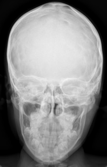
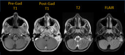
| Findings: Skull Radiograph: There is a destructive lesion in the left mastoid region. MRI of the Brain: There is a multilobulated lesion in the left mastoid with avid enhancement and significant bony destruction.
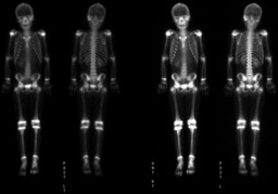
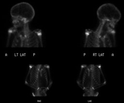
Whole Body Bone Scintigraphy: There is increased tracer uptake in the left mastoid which corresponds to the lesion on MRI. |
| DDx: Differential considerations include: Rhabdomyosarcoma Langerhans' cell histiocytosis Metastasis Lymphoma |
| Diagnosis: An excisional biopsy of a postauricular lymph node as well as a left temporal bone biopsy revealed Langerhans' cell histiocytosis. |
| References: Saliba I, Sidani K, El Fata F, Arcand P, Quintal MC, and Abela A. Langerhans' cell histiocytosis of the temporal bone in children. International Journal of Pediatric Otorhinolaryngology. Volume 72. Issue 6. Pages 775-786. |
| Comments: No comments posted. |
| Additional Details:
Case Number: 372671 The reader is fully responsible for confirming the accuracy of this content. |