Return to Case List with Diagnoses or Case List as Unknowns

METASTATIC OSTEOSARCOMA
Authored By: Keith Fischer and Dhanashree Rajderkar.
Patient: 50 year old male
History: 50 year old male with recent diagnosis of left distal ulnar mass, presumed to be a malignant lesion. Evaluate for bony metastasis.
Image Size:[small][as-submitted]

Fig. 1

Fig. 2

Fig. 3

Fig. 4

Fig. 5
Image Size:[small][as-submitted]

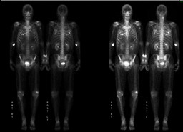
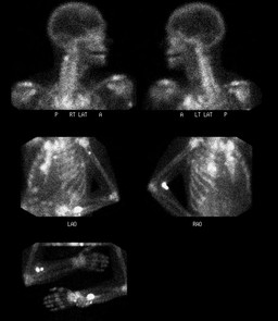
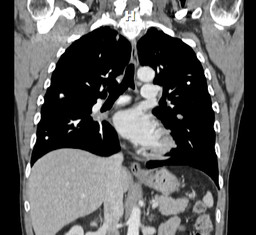
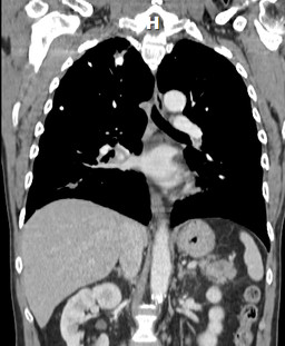
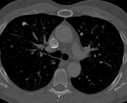
Findings:

Bone Scintigraphy: There is increased focal uptake in the left distal ulna, just proximal to the expanded distal expansile lesion. Focal uptake is also identified in the left proximal humerus. Focal increased activity in the region C2 vertebral body on the left side, could be in the pedicle.

Numerous foci of increased activity are seen scattered throughout the lung parenchyma on both sides, more on the right side. Increased activity is also identified in the right hilar region, which is thought to be a lymph node metastasis.

CT scan of Chest: Innumerable bilateral calcified and noncalcified pulmonary nodules. The appearance of these pulmonary nodules is most consistent with metastatic osteosarcoma versus chondrosarcoma.

DDx:

1. Ossifying metastases from the distal ulnar mass.

2.Calcified metastases from unknown primary.

3. Calcified nodules from old granulomatous disease.


Diagnosis: Ossifying metastases from the left distal ulnar mass.
General Discussion:

Most osteosarcomas occur in children and young adults in the metaphysis of a long bone especially around the knee joint. Multicentric osteosarcomas are caused by metastatic spread, although sometimes multiple synchronous tumors occur.

Metastatic spread of the tumor may be widespread, and occasionally ossifying metastases occur, especially in the lungs and lymph nodes. At diagnosis, 80% of patients have localized disease, while distant metastases can be identified in only 10%. The most common site of relapse in osteosarcoma has been in the lung. The pathological features of 66% of metastatic osteosarcoma of lung are essentially the same as the primary tumour. Lung metastases from osteosarcoma have been occasionally associated with lesions in bone, brain, chest wall, lymph nodes, and skin.

Calcification in metastatic pulmonary nodules, though unusual, may be seen in osteosarcoma and chondrosarcoma due to bone formation within the nodules. Dystrophic calcification may be seen in pulmonary metastases from giant cell tumours, papillary carcinoma of thyroid, synovial sarcomas, or treated metastatic disease. Mucinous adenocarcinomas of gastrointestinal tract and breast may also show calcified pulmonary metastases due to mucoid calcification.

Various findings have been reported in the metastatic disease of the lung in osteosarcoma, including solitary or multiple ossified parenchymal masses, ossified mediastinal lymph nodes, pleural calcifications, diaphragmatic deposits, pneumothorax, esophageal-mediastinal fistula, lymphangitis carcinomatosis, and pulmonary artery tumor.

Pleural deposits have been infrequently reported. Moreover, as hematogenous spread is the usual route of dissemination in osteosarcoma, metastatic lymph node involvement is uncommon.

Cavitation of metastatic nodules has also been described in osteosarcoma and necrosis of subpleural metastases may lead to formation of bronchopleural fistula and pneumothorax in 5% to 7% of cases.

References:

1.Rajendra Kumar, Ruppert David, John E. Madewell, and Marvin M. Lindell, Jr.,Radiographic spectrum of osteosarcoma; AJR 148:767-772, April 1987.

2. J-T LIN, MD, C-C YEN, MD, W-S WANG, MD, T-J CHIOU, MD and J-H LIU, MD and H-T WU, MD and P-M CHEN, MD; Case report: Unusual peritoneal spreading by metastatic osteosarcoma of the tibia; The British Journal of Radiology, 76 (2003), 337–338. 

3. Veenu Singla, Vivek Virmani, Rakesh Kapoor, Ritesh Agarwal and Niranjan Khandelwal, Massive Ossified Lung Masses; Indian J Chest Dis Allied Sci 2009;51:233-235. 

4. Tjeerd Zwaga, MD , Judith V. M. G. Bove´e, MD, PhD , Herman M. Kroon, MD, PhD;Best Cases from the AFIP: Osteosarcoma of the Femur with Skip Lymph Node, and Lung Metastases; RadioGraphics 2008; 28:277–283

Comments:
No comments posted.
Additional Details:

Case Number: 324525Owner(s): Keith Fischer and Dhanashree RajderkarLast Updated: 02-07-2013
Anatomy: Other   Pathology: Other
Modality: CT, Nuc MedAccess Level: Readable by all users, writable by NucMed Certifiers

Case has been viewed 19 times.
Certified by Keith Fischer on 02-04-2013

The reader is fully responsible for confirming the accuracy of this content.
Text and images may be copyrighted by the case author or institution.