Return to Case List with Diagnoses or Case List as Unknowns

BENIGN BONE TUMOR ON BONE SCINTIGRAPHY
Authored By: Farrokh Dehdashti and Archana Kantawala.
History: 62 year old man with a diagnosis of prostate cancer. The study is being done to evalaute for osseus metastatic disease.The patients serum PSA at the time of the study was 23.7 ng/mL
Image Size:[small][as-submitted]

Fig. 1

Fig. 2

Fig. 3

Fig. 4

Fig. 5
Image Size:[small][as-submitted]

Findings:

Bone scintigraphy -There is an area of mildly increased uptake in the distal diaphysis of the left femur. In addition, there are areas of abnormal uptake in the posterior aspect of the mid cervical spine, as well as the right lateral aspect of L4-L5, which are likely degenerative. The sacrum is not completely visualized due to activity in the bladder and patient's Foley catheter.

Plain films- Benign appearing lesion with chondroid matrix in the distal left femoral diaphysis corresponding to the area of tracer uptake on today's bone scan, consistent with an enchondroma.


General Discussion:

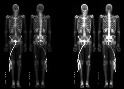
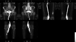
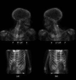
Bone metastases are often multiple at the time of diagnosis. In adults, the lesions generally occur in the axial skeleton and other sites with residual red marrow, although the lesions may be found anywhere in the skeletal system. Common sites for metastases are the vertebrae, pelvis, proximal parts of the femur, ribs, proximal part of the humerus, and skull. More than 90% of metastases are found in this distribution.

Bone scans are helpful in detecting these lesions, especially when they occur in sites that are difficult to evaluate on radiographs.

At initial presentation with prostate cancer, the value of a bone scan is limited in patients with a Gleason score of less than 7 and a prostate-specific antigen (PSA) level of less than 20 ng/mL. Those with a Gleason grade of greater than 6 may be candidates for a bone scan, irrespective of their PSA level. A bone scan may be performed as a baseline for treatment response in patients with recurrent metastatic disease at high risk of having bony metastatic disease.

Regardless of these guidelines, a bone scan is indicated in patients with prostate cancer who have symptoms suggesting bony metastases. Activity in the bone scan may not be observed until 5 years after micrometastasis has occurred; therefore, negative bone scan results do not rule out metastasis.

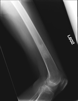
In addition to metastatic disease, benign bone lesions show a wide range of activity. Osteoblastomas, osteoid osteomas, chondroblastomas and giant cell tumors usually show intense activity.Enchondromas and Chondroblastomas show a moderate amount of increased activity. Fibrous cortical defects and nonossifying fibromas are usually mild or isointense compared to normal bone. Bone cysts are cold centrally but may have a warm rim due to increased bone remodelling.


References:

Essentials of nuclear medicine imaging. Fifth edition. Fred A. Mettler, Mitlon J. Guiberteau

http://emedicine.medscape.com/article/387840-overview#a19

http://emedicine.medscape.com/article/458011-overview

Comments:
No comments posted.
Additional Details:

Case Number: 323766Owner(s): Farrokh Dehdashti and Archana KantawalaLast Updated: 02-07-2013
Anatomy: Skeletal System   Pathology: Benign Mass, Cyst
Modality: Conventional Radiograph, Nuc MedAccess Level: Readable by all users, writable by NucMed Certifiers
Keywords: bsnm

Case has been viewed 25 times.
Certified by Farrokh Dehdashti on 11-09-2011

The reader is fully responsible for confirming the accuracy of this content.
Text and images may be copyrighted by the case author or institution.