|
| Patient: 36 year old male |
| History: 36 year old male: with primary sclerosing cholangitis, 3 days status post orthotopic liver transplant who presents with elevated transaminases. Abdominal sonogram performed a day prior showed a fluid collection in the porta hepatis. There was a clinical concern for biliary leak. |
Image Size:
|
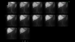
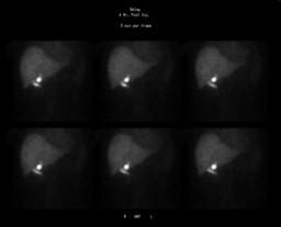
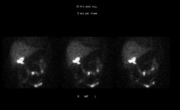
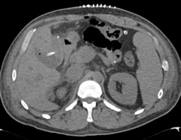
| Findings: Hepatobiliary Scintigraphy: Findings: There is delayed accumulation of the tracer by the liver and persistent blood pool activity, consistent with moderately decreased hepatic function. Delayed images show a loculated collection of activity that develops along the inferior margin of the liver, in the region of the porta hepatis, and correlates with the collection identified on ultrasound. 4-5 hour delayed images show a second loculated collection, which is more linear and may represent pooling of tracer or a small amount of tracer entering the bowel. Obvious flow of tracer into the bowel is not identified. Additional images were obtained approximately 20 hours after injection of tracer. The small linear focus previously seen immediately inferior to the gallbladder fossa now can be seen to extend further, and is now consistent with excretion of tracer into bowel. Additional bowel activity is also seen. Impression: 1. Delayed tracer uptake and persistent blood pool activity, consistent with moderate hepatic dysfunction. 2. Loculated collection along the inferior margin of the liver near the porta hepatis, which correlates with the collection seen on ultrasound and is most consistent with a small loculated biloma. CT Scan: Impression: 1. Status post recent liver transplant. 2. A 6.6 x 5.4 x 5.4 cm fluid in the gallbladder fossa compatible with the biloma. |
| DDx: 1.Normal gallbladder (unlikely in this patient s/p cholecystectomy at time of liver transplant). 2.Biloma. |
| Diagnosis: Biloma due to iatrogenic injury to Duct of Luschka proven after exploratory laparotomy. |
| References: 1.Albishri et al; Bile leak from Duct of Luschka after liver transplantaion; Brief communications:Clinical Transplantation;Transplantation:27 July 2001-Vol 72,Issue 2,pg 338-340. 2.J. M. Ramia, PhD, K. Muffak, MD, A. Mansilla, PhD, J. Villar, PhD, D. Garrote, PhD, and J. A. Ferron, PhD; Postlaparoscopic cholecystectomy bile leak secondary to an accessory duct of Luschka. JSLS. 2005 Apr-Jun;9(2):216-7. 3.K. Sharif and J. de Ville de Goyet; Bile Duct of Luschka Leading to Bile Leak After Cholecystectomy—Revisiting the Biliary Anatomy; J Pediatr Surg 38:E60. |
| Comments: No comments posted. |
| Additional Details:
Case Number: 318486 The reader is fully responsible for confirming the accuracy of this content. |