Return to Case List with Diagnoses or Case List as Unknowns

METASTATIC OSTEOSARCOMA WITH ASSOCIATED SOFT TISSUE COMPONENT
Authored By: Keith Fischer and Kenneth Winkler.
Patient: 11 year old female
History: 11 year old girl, with lower extremity pain.
Image Size:[small][as-submitted]

Fig. 1
Bone Scintigraphy

Fig. 2
Bone Scintigraphy

Fig. 3
Femur Radiography

Fig. 4
Femur Radiography

Fig. 5
Chest Computed Tomography

Fig. 6
Chest Computed Tomography
Image Size:[small][as-submitted]

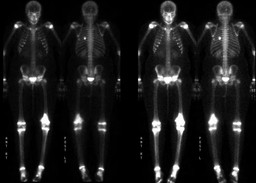
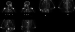
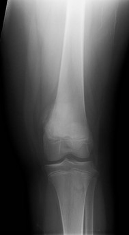
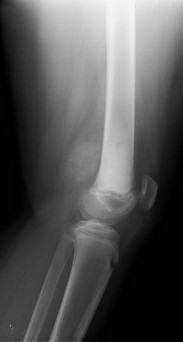
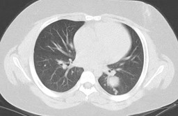
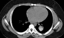
Findings:

FINDINGS (Bone Scintigraphy):

RADIOPHARMACEUTICAL: 16.7 mCi Tc-99m MDP i.v.


Delayed whole-body scintigrams were obtained. No prior study is available for comparison.


There is markedly increased radiotracer uptake at the distal end of the left femur. A faint focus of radiotracer uptake is seen at the left lateral aspect of the L2 vertebral body, best appreciated on the posterior images. Additionally there is a faint focus of increased radiotracer uptake in possibly the L3 vertebral body, appreciable only on the anterior views. There is a focus of increased radiotracer uptake located in the left hemithorax, between the ribs, best seen on the posterior views . Lateral to this is another fainter focus of increased radiotracer uptake, a little below the inferior tip of the left scapula.


FINDINGS (Radiography):

Radiographs of the left femur are submitted for interpretation without prior studies for comparison. There is a soft tissue mass with increased density in the posterior and distal left leg. There is sclerosis of the distal left femur at the metaphysis. Minimal periosteal reaction is seen. No fracture or dislocation is identified.


FINDINGS (Computed Tomography):

There are numerous pulmonary nodules scattered throughout all lobes of left and right lung, left greater than right. The 2 largest pulmonary nodules in the lingula and left lower lobe contain calcifications. These measure 2 cm x 1.8 cm and 3.6 cm x 3.2 cm, respectively.

Diagnosis: Metastatic osteosarcoma, including osseous and pulmonary metastasis.
General Discussion:

HISTORY:

11 year old girl, with initial presentation of lower extremity pain.  Patient was found to have a distal left femoral lesion on radiography with subsequent biopsy demonstrating osteosarcoma.  Additional work-up, including bone scintigraphy and CT chest demonstrated osseous and pulmonary metastasis.


DISCUSSION:

Differential Diagnosis for Multifocal Disease on Osseous Scintigraphy

Metastatic Disease

Degenerative Change

Trauma

Metabolic Osseous Disease

Paget's

Infection


Differential Diagnosis for Localized Soft Tissue Lesions

Soft Tissue Tumors

Tumoral Calcinosis

Myositis Ossificans

Tissue Infarction

Polymyositis

Calcific Tendonitis


Osteosarcoma is the eighth most common malignancy in children.  Patients typically present with pain, although larger lesions may present with swelling.  The lesions can also weaken the underlying bone, which could result in a pathologic fracture and initial presentation.  Diagnosis usually begins with radiographs, followed by additional imaging as needed, with subsequent surgical biopsy.  The current treatment stragedy involves neoadjuvant chemotherapy followed by surgical resection, to include a limb salvage procedure if possible.  Prognosis ranges between 30-90%, depending on staging.  Given the widespread pulmonary metastatic disease, this patient would be a stage III, which is associated with a poorer prognosis.

References: Mettler, Fred and Milton Guiberteau. Essentials of Nuclear Medicine Imaging. 5th ed. Philadelphia, PA: Saunders Elsevier, 2006, pp 243-292.
Comments:
No comments posted.
Additional Details:

Case Number: 301748Owner(s): Keith Fischer and Kenneth WinklerLast Updated: 02-07-2013
Anatomy: Skeletal System   Pathology: Neoplasm
Modality: CT, Conventional Radiograph, Nuc MedAccess Level: Readable by all users, writable by NucMed Certifiers
Keywords: bsnm; 4.322

Case has been viewed 19 times.
Certified by Keith Fischer on 06-10-2011

The reader is fully responsible for confirming the accuracy of this content.
Text and images may be copyrighted by the case author or institution.