Return to Case List with Diagnoses or Case List as Unknowns

MCCUNE-ALBRIGHT SYNDROME
Authored By: Bennett Greenspan and Livnat Uliel.
Patient: 6 year old male
History: 7 y-o boy with suspected precocious puberty.
Bone scan is requested as part of the workup.
Image Size:[small][as-submitted]

Fig. 1
Figure 1

Fig. 2
Figure 2

Fig. 3
Figure 3

Fig. 4
Figure 4
Image Size:[small][as-submitted]

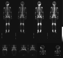
Findings:

Presented whole body scan and spot images of the pelvis and skull (Figure 1).
Radiopharmaceutical : 5.8 mCi Tc-99m MDP IV
Moderately increased activity is seen in the left frontal bone and medial aspect of the left femoral neck.


Bone survey was performed. The relevant images are presented.


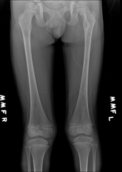
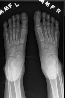
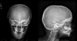
There is a radiolucent lesion with peripheral increased density lesion in the medial aspect of the femoral neck and proximal diaphysis. Additional less defined mixed sclerotic and lucent lesions are seen in the distal left femur (Figure 2) and left first metatarsal bone (Figure 3).

A ground glass lesion with bone expansion is present in the left frontal bone (Figure 4). The findings likely represent fibrous dysplasia and are in correlation with the scintigraphic findings.


Additional information:  On physical exam a café-au-lait macule is present on the left leg and a 10 cm hyperperpigmented lesion on the upper part of the back extending to the neck, with irregular borders.


DDx: In this case the clinical presentation and imaging suggest McCune-Albright Syndrome
In general differential diagnosis of FD include Paget disease (in older age group), Osteogenesis Imperfecta (usually present with deformed long bones with moderate diaphyseal uptake) and bone tumors (primary and metastasis).
Diagnosis:


Diagnosis:  McCune-Albright Syndrome

General Discussion:


Fibrous dysplasia (FD) of bone is a genetic, non-inheritable disease, caused by post-zygotic mutation in the gene coding for the guanine nucleotide binding, alpha stimulating (GNAS) complex.
McCune-Albright Syndrome (MAS) is the most severe form of the mutation described as a triad of polyostotic unilateral FD, endocrine dysfuction and cafe´-au-lait cutaneous spots, and represents less than 5% of patients with FD.  Patients with polyostotic FD often have renal phosphate wasting with various degrees of hypophosphataemia. The dermatologic findings are describe as coast of Maine café-au-lait spots which are yellowish to brownish patches of cutaneous pigmentation with irregular border (as oppose to coast of California spots of neurofibromatosis). The diagnosis can be established by imaging, molecular biology and histology.

Clinical presentation of FD are bone pain that might be associated with limp, bone deformities and fractures. Involvement of the skull may narrow the neural foramina, cause obliteration of the sinuses or displace the orbit.  The common location is rib cage (30%), craniofacial bones (25%), femoral neck and tibia (25%) and pelvis.  Lesions are usually extend from the medullary cavity to the cortex, are fibrous or cystic in nature, causing radiolucent or ground glass appearance and expansion of the bone. Usually there is well defined sclerotic margin of reactive bone. Classic radiologic appearance is endosteal scalloping with thinned cortex and intervening normal cortex

Bone scan can help in searching for all affected bones and therefore to differentiate between monostotic and polyostotic disease.  Increased activity is usually seen during early perfusion and on delayed images, but the uptake is variable. Significant increased activity on bone scan is also seen in pathologic fractures and malignant degeneration and usually associated with acute pain.

References:

References:

Diagnostic Imaging Nuclear Medicine, Morton and Clark, First Edition, AMIRSYS

Chapurlat RD, Orcel P, Fibrous Dysplasia of Bone and McCune-Albright Syndrome. Best Practice and Research Clinical Rheumatology 2008;22:55-69

Radiology Review Manual, Dahnert, Fourth Edition, LWW

Comments:
No comments posted.
Additional Details:

Case Number: 300084Owner(s): Bennett Greenspan and Livnat UlielLast Updated: 02-07-2013
Anatomy: Skeletal System   Pathology: Other
Modality: Conventional Radiograph, Nuc MedAccess Level: Readable by all users, writable by NucMed Certifiers
Keywords: bsnm, mccune-albright syndrome, fibrous dysplasiaACR: 40000.85200

Case has been viewed 34 times.
Certified by Bennett Greenspan on 09-24-2011

The reader is fully responsible for confirming the accuracy of this content.
Text and images may be copyrighted by the case author or institution.