Return to Case List with Diagnoses or Case List as Unknowns

VENOUS COLLATERALS SECONDARY TO SUPERIOR VENA CAVAL OBSTRUCTION
Authored By: kwinkl01 and Barry Siegel, Prof of Radiology.
Patient: 59 year old female
History:

HISTORY:

59-year-old woman with peptic ulcer disease with an associated jejunal ulcer and recent hematochezia and hypotension.  Evaluate for site of active bleeding.

Image Size:[small][as-submitted]

Multimedia: 298646_1_submitted.avi
Radionuclide Angiography

Multimedia: 298646_2_submitted.avi
Sequential Anterior Scintigraphy

Fig. 3
Computed Tomography
Image Size:[small][as-submitted]

Findings:

RADIOPHARMACEUTICAL: 27.1 mCi Tc-99m in vitro labeled red cells i.v.


 FINDINGS (SCINTIGRAPHY):

Sequential anterior and posterior abdominal images were obtained through 60 minutes (Additional anterior images, not shown, were obtained through 90 minutes. The radionuclide angiographic phase demonstrates extensive venous collaterals. There is an attenuation artifact overlying the spleen on the anterior images.  No abnormal foci of labeled red cell extravasation are seen.


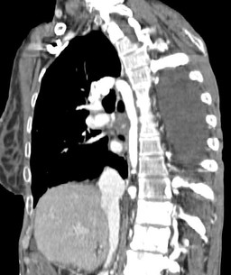
FINDINGS (COMPUTED TOMOGRAPHY):

A single oblique coronal computed tomographic image demonstrates marked enlargement of the azygos venous system.  There is obstruction of the superior vena cava just inferior to the azygos arch.

Diagnosis:

No evidence for active gastrointestinal bleeding. 


Extensive venous collaterals compatible with known superior vena cava obstruction. 


Attenuation artifact overlying the spleen on the anterior images attributable to a telemetry monitor.

General Discussion:

HISTORY: 

59-year-old woman with peptic ulcer disease with an associated jejunal ulcer and recent hematochezia abd hypotension.  Evaluate for site of active bleeding.  She has a known superior vena cava obstruction just inferior to the azygos arch.  She also has pancreatic cancer, status post resection, chemotherapy, and radiation.


DISCUSSION:

Superior vena cava syndrome usually results from direct compression of the superior vena cava secondary to adjacent masses.  The most common cause of superior vena cava obstruction is bronchogenic carcinoma.  Other malignancies, such as lymphoma, can also cause superior vena cava obstruction.  Primary mediastinal masses, such as those of thymic origin, can also result in obstruction of the superior vena cava.  Thrombosis, usually secondary to intravascular devices, is the most common non-compressive etiology of superior vena cava syndrome.  Patients usually present with shortness of breath, as well as facial and/or upper extremity swelling.  Diagnosis is typically confirmed with contrast enhanced computed tomography, although conventional angiography or magnetic resonance angiography could also be utilized.  Treatment usually involves surgical resection, medical therapy, usually in the form of steroids, to decrease the amount of inflammation, or radiation to decrease the size of the obstructing mass.  Symptomatic treatment often involves diuretics to decrease the amount of swelling.  Prognosis in cases where tumor is the cause is poor, as superior vena cava syndrome typically indicates an advanced stage of malignancy.

Comments:
No comments posted.
Additional Details:

Case Number: 298646Owner(s): kwinkl01 and Barry Siegel, Prof of RadiologyLast Updated: 02-07-2013
Anatomy: Vascular/Lymphatic   Pathology: Vascular
Modality: CT, Nuc MedAccess Level: Readable by all users, writable by NucMed Certifiers
Keywords: ginm

Case has been viewed 23 times.
Certified by Barry Siegel on 03-16-2011

The reader is fully responsible for confirming the accuracy of this content.
Text and images may be copyrighted by the case author or institution.