Return to Case List with Diagnoses or Case List as Unknowns

PATHOLOGIC FRACTURE
Authored By: kwinkl01 and Keith Fischer.
Patient: 68 year old female
History: HISTORY: 68-year-old woman with hypercalcemia with persistent right upper extremity pain and swelling.
Image Size:[small][as-submitted]

Fig. 1
bone scintigraphy

Fig. 2
radiography
Image Size:[small][as-submitted]

Findings:

RADIOPHARMACEUTICAL: 21.6 mCi Tc-99m MDP i.v.


FINDINGS (Scintigraphy): Delayed whole-body scintigrams were obtained. There is marked swelling of the right upper extremity and right chest wall, likely representing lymphedema versus possible swelling secondary to venous thrombosis. Adjacent to the proximal right humerus, there is a region of decreased uptake in the soft tissues. In the proximal right humerus, there is moderate uptake which is less prominent than would be expected given the age of the patient's fracture. This represents poor callus formation versus possible tumor involvement. In the right humerus distal to this area of moderate uptake, there is a focus of decreased uptake. There are no additional abnormal areas of increased or decreased uptake identified.


FINDINGS (Radiography): There is a comminuted ununited fracture through the proximal right humerus with distraction and posterior angulation. No other fractures are identified. There is no shoulder dislocation. There is marked edema of the right arm. Extensive osteopenia is seen throughout the humerus.

Diagnosis: Pathologic fracture secondary to large B cell lymphoma.
General Discussion:

FULL HISTORY:

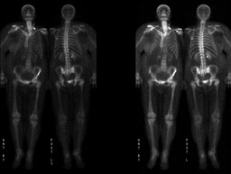
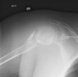
68-year-old woman with history of comminuted right humeral fracture eight months prior. She now presents with hypercalcemia and persistent right upper extremity pain along with swelling of the right upper extremity and right chest wall. A recent chest CT demonstrates a comminuted fracture of the proximal right humerus with a large adjacent fluid collection with possible soft tissue mass component. The patient has no known history of cancer.  Evaluate for primary or metastatic disease.


DIFFERENTIAL DIAGNOSIS: Localized bone scintigraphy uptake (Mettler, 2006)

trauma

degenerative changes (arthritis)

infection

primary bone tumor (benign and malignant)

metastatic disease

Paget's disease, fibrous dysplasia

hyperemia

overlying soft tissue activity

decreased overlying soft tissue (decreased attenuation)


DISCUSSION:

Pathologic fractures occur in bone that has been weakened secondary to a disease process, usually osteoporosis, although primary and secondary bone lesions, to include both benign and malignant etiologies can result in pathologic fractures.  Pathologic fractures can occur during normal activity or minor trauma secondary to the weakening of the underlying bone.  Pathologic fractures represent a serious co-morbidity in patients with osseous metastatic disease.  Prevention of pathologic fractures by utilizing proactive treatments can result in better patient outcomes, lower cost, and less difficult operative procedures.  Therefore, it is important to determine which lesions may require preventative treatment.  Many characteristics can be used for determining the risk of pathologic fracture including type of cancer, type of treatment, size of the lesion, location of the lesion, characteristics of the lesion and symptoms due to the lesion.

References: Mettler, Fred and Milton Guiberteau. Essentials of Nuclear Medicine Imaging. 5th ed. Philadelphia, PA: Saunders Elsevier, 2006, pp 244.
Comments:
No comments posted.
Additional Details:

Case Number: 298403Owner(s): kwinkl01 and Keith FischerLast Updated: 02-07-2013
Anatomy: Skeletal System   Pathology: Neoplasm
Modality: Conventional Radiograph, Nuc MedAccess Level: Readable by all users, writable by NucMed Certifiers
Keywords: bsnm

Case has been viewed 17 times.
Certified by Keith Fischer on 06-10-2011

The reader is fully responsible for confirming the accuracy of this content.
Text and images may be copyrighted by the case author or institution.