Return to Case List with Diagnoses or Case List as Unknowns

VENTILATION-PERFUSION SCINTIGRAPHY
Authored By: Keith Fischer and Livnat Uliel.
Patient: 77 year old male
History:

77 year old man admitted with worsening shortness of breath.

His past medical history includes: severe COPD, paroxysmal atrial fibrillation, and ulcerative colitis.

Image Size:[small][as-submitted]

Fig. 1
Ventilation-Perfusion Scintigraphy

Fig. 2
PA and Lateral Chest X-rays

Fig. 3
Clinical Prediction Rules for Pulmonary Embolism
Image Size:[small][as-submitted]

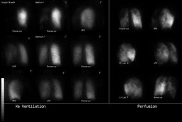
Findings:

The ventilation-perfusion scintigraphy and PA and Lateral chest x-rays performed the same day are presented

Ventilation-Perfusion Scan (Fig. 1)

Radiopharmaceuticals: 17.2 mCi Xe-133 gas by inhalation and 4.7 mCi Tc-99m MAA i.v.


The Xe-133 ventilation images show markedly diminished ventilation of the left lung on the initial breath and wash-in images. There is a focal ventilation defect in the initial breath image in the right lung apex which subsequently fills in on the wash-in images.   Both the lungs are hyper-inflated with significant retention of Xe-133 during the washout phase bilaterally.
The perfusion images show diffuse decrease in the left lung perfusion.  There are patchy nonsegmental perfusion defects bilaterally.  There is a small focal perfusion defect in the right lung apex which is matched by the ventilation defect. No moderate or large segmental mismatches are identified that suggest pulmonary embolism.

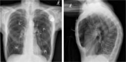
PA and lateral views of the chest (Fig. 2)

The heart is normal in size.  Single lead left transvenous pacer lead extends to the right ventricle.
There are severe emphysematous changes bilaterally, with no evidence of focal consolidation or mass lesion. No pleural effusion or pneumothorax seen. Pulmonary arterial enlargement is present.
Mild compression deformities of a lower and midthoracic vertebral bodies.

DDx:


Low likelihood ratio for pulmonary embolism

Diagnosis:

No Pulmonary Embolism
Chronic obstructive pulmonary disease exacerbation

The patient also underwent Doppler ultrasound of the lower extremities with no evidence of DVT

General Discussion:

The diagnosis of low likelihood probability, ≤20% likelihood of acute PE, was based on the following criteria: small perfusion defects which are nonsegmental and matched V-P defects with clear chest radiograph. 
The scan result should be integrated with the clinical pre-test likelihood  based on clinical prediction rules for pulmonary embolism, such as Well’s criteria, Geneva score or the revised Geneva score, which all demonstrate similar accuracy (Ceriani et al., Fig 3). 

In this case we don’t have all the clinical data to determine the pre-test  likelihood, nevertheless in cases of low pre-test clinical likelihood, no further study or workup is needed.  In case of intermediate or high pre-test clinical likelihood  venous Doppler ultrasound or pulmonary CTA may be useful to make the final diagnosis. The Doppler ultrasound of the lower extremities was negative for DVT in this case and therefore helps to rule out acute PE.


It should be mentioned that in hemodynamically stable patients with low/intermediate clinical probability for PE and normal D-dimer value (sensitive enzyme-linked immunosorbent assay), no further workup in needed (Agnelli and Becattini).  Recent systematic review demonstrated that negative VIDAS D-dimer (enzyme-linked fluorescent immunoassay) in combination with a non-high pretest probability can safely exclude pulmonary embolism with negative predictive value of 99.9% (Carrier M et al.).  The use of the D-dimer is of limited value in patients with high clinical probability of pulmonary embolism.  Increased level of D-dimer in cancer patients, pregnant women, hospitalized and elderly patients, reduce the specificity of the test (Agnelli and Becattini). 


References:

Diagnostic Imaging Nuclear Medicine, Morton and Clark, First Edition, AMIRSYS

Carrier M et al. VIDAS D-dimer in Combination with Clinical Pre-test Probability to Rule Out Pulmonary Embolism, A Systematic Review of Management Outcome Studies. Thromb Haemost 2009;101:886-892

Agnelli and Becattini. Acute Pulmonary Embolism. NEJM 2010;363:266-74

Ceriani E et al. Clinical Prediction Rules for Pulmonary Embolism: A Systematic Review and Meta-Analysis. J Thromb Haemost 2010;8:975-70

Comments:
No comments posted.
Additional Details:

Case Number: 293895Owner(s): Keith Fischer and Livnat UlielLast Updated: 02-07-2013
Anatomy: Cardiopulmonary   Pathology: Other
Modality: Conventional Radiograph, Nuc MedAccess Level: Readable by all users, writable by NucMed Certifiers
Keywords: vqnmACR: 60000.72000

Case has been viewed 13 times.
Certified by Keith Fischer on 01-23-2013

The reader is fully responsible for confirming the accuracy of this content.
Text and images may be copyrighted by the case author or institution.