Return to Case List with Diagnoses or Case List as Unknowns

INDIUM-111 WBC SCINTIGRAPHY IN A CASE OF LEFT VENTRICULAR ASSISTED DEVICE INFECTION
Authored By: Garima Agrawal and Keith Fischer.
Patient: 61 year old male
History: 61 year old male: who has had placement of a left ventricular assist device for known cardiomyopathy     
Image Size:[small][as-submitted]

Fig. 1

Fig. 2

Fig. 3

Fig. 4

Fig. 5
Image Size:[small][as-submitted]

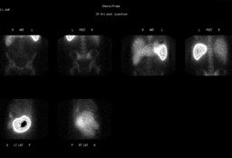
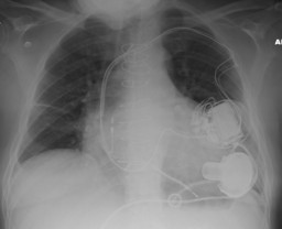
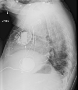
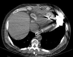
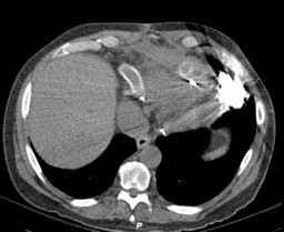
Findings:


In-111 WBC scintigraphy: There is a focal area of increased radiotracer uptake in the pocket of the left ventricular device in the left anterior chest. There is a small linear focus of activity medial to the device, which could be in the proximal tubing. No definite activity is identified along the drive line in the abdomen.


Chest Radiograph: Left ventricular assist device is seen. There is a 2-lead left transvenous pacemaker defibrillator with leads in the right atrium and right ventricle. Median sternotomy wires and clips from coronary bypass procedure are seen. There is a coronary artery stent in place. Right base atelectasis present.


Chest CT: There is fluid and mild stranding adjacent to the proximal and distal aspects of the left ventricular assist device
driveline.

DDx:

LVAD drive line infection

LVAD device infection

Diagnosis: Infection of the pocket of left ventricular assist device with possible extension into the tubing leading to the heart.
General Discussion:


Implantation of a left-ventricular-assist device (LVAD) is increasingly becoming the therapy of choice in patients with advanced heart failure. However, device-related infection continues to be a morbid problem and remains a serious limitation to long-term use of the device  

Infection and sepsis have a high prevalence and remain a major cause of morbidity and mortality in patients with LVAD. Major clinical issues include diagnosing the infection early, determining the location and extent of the infection, and assessing treatment efficacy.

The use of In-111 labeled WBC's is an efficient means of detecting the extent of the infection involving the LVAD

Specific Discussion:


Leukocyte planar scintigraphy has a high sensitivity (98%) and specificity (89%) for identifying vascular graft infection [1-3]. A recent study by Litzler et. al. demonstrated the feasibility and potential usefulness of radiolabeled-leukocyte SPECT/CT in the management of infection in LVAD patients. They concluded that leukocyte SPECT/CT is able to determine the precise anatomic location and extent of a suspected infection and could lead to improved therapeutic strategies [4].

References:


  1. Burroni L, D’Alessandria C, Signore A. Diagnosis of vascular prosthesis infection: PET or SPECT? J Nucl Med. 2007;48:1227–1229.
  2. Becker W, Meller J. The role of nuclear medicine in infection and inflammation. Lancet Infect Dis. 2001;1:326–333.
  3. Roach PJ, Bailey DL. Combining anatomy and function: the future of medical imaging. Intern Med J. 2005;35:577–579
  4. Litzler PY, Manrique A, Etienne M. Leukocyte SPECT/CT for detecting infection of left-ventricular-assist devices: preliminary results. J Nucl Med. 2010 Jul;51(7):1044-8. Epub 2010 Jun 16.
Comments:
No comments posted.
Additional Details:

Case Number: 285372Owner(s): Garima Agrawal and Keith FischerLast Updated: 02-07-2013
Anatomy: Cardiopulmonary   Pathology: Infection
Modality: CT, Nuc MedAccess Level: Readable by all users, writable by NucMed Certifiers
Keywords: lvad, iwnm, in-111 leukocyte scintigraphy

Case has been viewed 20 times.
Certified by Keith Fischer on 02-04-2013

The reader is fully responsible for confirming the accuracy of this content.
Text and images may be copyrighted by the case author or institution.