Return to Case List with Diagnoses or Case List as Unknowns

ILEAL CONDUIT URINARY LEAK
Authored By: luliel01 and Barry Siegel, Prof of Radiology.
Patient: 60 year old male
History:

60 year-old man with extensive local recurrence of rectal cancer after low anterior resection and chemoradiation.  He underwent a revision abdominal perineal resection and radical cystoprostatectomy with ileal conduit diversion. The surgery was complicated by delayed abdominal and perineal wound healing which progressed to drainage from the perineal wound.  Analysis of fluid drained from the perineum revealed a high creatinine level suggesting urine leak.


For further evaluation of the renal function and ntegrity of the ileal conduit,and for possible localization of the level of the leak, a renal scan was performed.

Image Size:[small][as-submitted]

Fig. 1
Fig. 1

Fig. 2
Fig. 2

Fig. 3
Fig. 3

Fig. 4
Fig. 4

Fig. 5
Fig. 5

Multimedia: 283225_6_submitted.avi
AVI File
Image Size:[small][as-submitted]

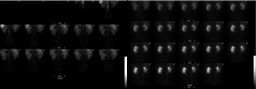
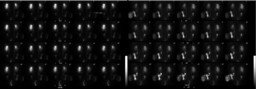
Findings:

Presented are the posterior flow and renal function images (Fig. 1) followed by posterior delayed images and anterior images after furosemide administration (Fig. 2).


Radiopharmaceutical: 8.3 mCi Tc-99m MAG3 I.V. and 40 mg furosemide I.V.


The posterior abdominal radionuclide angiogram demonstrates normal, symmetric perfusion of the kidneys. Sequential renal images show the kidneys to be of normal size with moderately enlarged collecting systems bilaterally, appearing as central photon-deficient areas on early images.  Both kidneys demonstrate prompt uptake of the radiopharmaceutical.  The left kidney demonstrates mildly delayed excretion into the collecting system, but the right kidney demonstrates markedly delayed excretion with only a small amount of radiopharmaceutical seen in the right renal collecting system after 20 minutes.


There is moderate retained activity in the left collecting system, which appears moderately enlarged.  There is mild retained activity in the right collecting system, which appears normal in size.


To evaluate for obstruction, the patient was given 40 mg furosemide via slow intravenous injection approximately 45 minutes after the start of the examination.  Sequential images were obtained for an additional 34 minutes (only the first 20 minutes are presented).  There is markedly delayed clearance of pelvicalyceal activity on the left after diuretic administration.  On the right, there is moderately delayed clearance of activity from the pelvicalyceal system.


After diuretic administration, the half-time of tracer clearance from the right kidney is 37 minutes and from the left kidney is 74 minutes (Fig. 3).


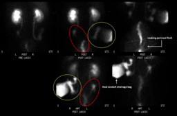
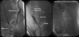
The dynamic urine flow to the ileal conduit on the right and to the perineal area on the left after furosemide administration can be appreciated on the attached cine (Fig. 6 - AVI File).

The left ureter does not appear to connect with the ileal conduit and radiopharmaceutical tracks into the pelvis, most likely representing a urine leak (Fig. 4, red ellipses, best visualized on the anterior view).  During this study, perineal fluid leaked onto the imaging table.  This perineal fluid was found to be radioactive, further confirming the presence of a urine leak.  Radiopharmaceutical is seen draining from the right ureter into the ileal conduit (Fig. 4, green circles).

Diverting bilateral nephrostomies tubes were placed the day after.


Figure 5 shows images performed three weeks later during catheter exchange. The image on the left shows a catheter in the right ureter with the tip in the ileal conduit and contrast filling the right ureter and the ileal conduit. The image in the middle demonstrates a catheter in the left ureter with extravasation of contrast to the pelvis, similar to the findings on the renal scan. A nephrouretral stent to the ileal conduit was placed on the right, as seen on the right image, and a diverting nephrostomy was exchanged on the left.

Diagnosis: Urine leak that is most likely secondary to disrupted anastomotic site of the left ureter to the ileal conduit diagnosed on renal scan and confirmed on antegrade ureterography.
General Discussion:

Most of the literature on urinary leak is related to traumatic injury with CT being the imaging method of choice.  In the setting of trauma, renal scintigraphy also may be helpful in documenting the presence of a functioning kidney in a patient with a contraindication to use of iodinated contrast material or in follow-up after repair of renovascular trauma.  Most of the literature on scintigraphic imaging of urinary leak is related to its use after renal transplantation.  


In patients with impaired renal function, as in our case, when the use of iodinated contrast media is limited, renal scintigraphy can be helpful.  The dynamic acquisition can not only demonstrate the leak but also can localize the level of extravasation.

References:

Kawashima A, Sandler CM, Corl FM, West OC, Tamm EP, Fishman EK, Goldman SM.  Imaging of renal trauma: a comprehensive review. RadioGraphics 2001; 21:557–574

Titton RL, Gervais DA, Hahn PF, Harisinghani MG, Arellano RS, Mueller PR.  Urine leaks and urinomas: diagnosis and imaging-guided intervention.  RadioGraphics 2003; 23: 1133-1147

Comments:
No comments posted.
Additional Details:

Case Number: 283225Owner(s): luliel01 and Barry Siegel, Prof of RadiologyLast Updated: 02-07-2013
Anatomy: Genitourinary (GU)   Pathology: Other
Modality: Nuc Med, OtherAccess Level: Readable by all users, writable by NucMed Certifiers
Keywords: rsnmACR: 70000.32000, 80000.45300

Case has been viewed 20 times.
Certified by Barry Siegel on 03-16-2011

The reader is fully responsible for confirming the accuracy of this content.
Text and images may be copyrighted by the case author or institution.