Return to Case List with Diagnoses or Case List as Unknowns

HYPERTROPHIC OSTEOARTHROPATHY
Authored By: Bennett Greenspan and Dhanashree Rajderkar.
Patient: 60 year old male
History: 60 year old male: with back pain and left thigh pain. Rest of the history is withheld.
Image Size:[small][as-submitted]

Fig. 1

Fig. 2

Fig. 3

Fig. 4
Image Size:[small][as-submitted]

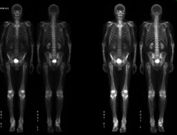
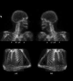
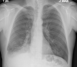
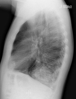
Findings:

Patient had undergone PET CT in an outside hospital a month ago

PET CT showed right lower lobar mass, and focal increased uptake in the left proximal femur.

Bone scan findings:

•Diffuse increased cortical uptake in bilateral upper and lower extremities, in a patient with known lung mass, most probably represents hypertrophic osteoarthropathy .

 •No mets.

Chest Radiograph.

Mass in the right lower lobe with adjacent consolidation/atelectasis.

DDx:

1.Paget's disease.

2.Acromegaly

3.Fibrous dysplasia.

4.Endosteal Hyperostosis.

5.Diffuse Idiopathic skeletal hyperostosis.

The appearance on this bone scan would not be typical for items 1-3.

Diagnosis: Pulmonary Hypertrophic osteoarthropathy.
General Discussion:

Pateint underwent right lower lobectomy.

8 cm mass, which was moderate-poorly differentiated adenocarcinoma was found.


Hypertrophic pulmonary osteoarthropathy

Isotope bone scanning with technetium-99m (99m Tc)–labeled diphosphonate shows evidence of hypertrophic pulmonary osteoarthropathy (HPOA) early in the course of disease; in addition, the sensitivity of isotope bone scans is greater than that of other imaging methods.

 Hypertrophic pulmonary osteoarthropathy (HPOA) usually involves diaphyseal and metaphyseal periostitis. Periosteal proliferation is usually single or laminated and is either regular or irregular.

Initially, periostitis is symmetric and involves the tibia, fibula, radius, ulna, and, less commonly, the femur, humerus, metacarpals, metatarsal, and phalanges on both sides. Eventually, periosteal proliferation extends into the metaphysis. In rare cases, periostitis affects the ribs, clavicles, and scapula.

Scintigraphic findings in patients with hypertrophic pulmonary osteoarthropathy (HPOA) appear earlier than radiographic findings do, and they correspond well to the clinical findings. Activity decreases with successful therapeutic measures, such as surgery or radiation therapy. Tumor recurrence may be associated with recurrent findings of increased radionuclide uptake.

References:

1.http://emedicine.medscape.com/article/390998-imaging

Imaging in Hypertrophic Osteoarthropathy: Imaging

Author: Ali Nawaz Khan, MBBS, FRCS, FRCP, FRCR, Consultant Radiologist and Honorary Professor, North Manchester General Hospital Pennine Acute NHS Trust, UK

2.Diagnostic imaging in hypertrophic osteoarthropathy. Clin Exp Rheumatol.  1992; 10 Suppl 7:27-33 

Comments:
No comments posted.
Additional Details:

Case Number: 279367Owner(s): Bennett Greenspan and Dhanashree RajderkarLast Updated: 02-07-2013
Anatomy: Skeletal System   Pathology: Metabolic
Modality: Conventional Radiograph, Nuc MedAccess Level: Readable by all users, writable by NucMed Certifiers
Keywords: hypertrophic osteoarthropathy

Case has been viewed 39 times.
Certified by Bennett Greenspan on 08-26-2011

The reader is fully responsible for confirming the accuracy of this content.
Text and images may be copyrighted by the case author or institution.