Return to Case List with Diagnoses or Case List as Unknowns

ACTIVE HETEROTOPIC OSSIFICATION
Authored By: kwinkl01 and Delphine Chen.
Patient: 48 year old female
History: 48-year-old woman with persistent left hip and right shoulder pain.
Image Size:[small][as-submitted]

Fig. 1
bone scintigraphy

Fig. 2
left hip radiograph

Fig. 3
right shoulder radiograph
Image Size:[small][as-submitted]

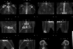
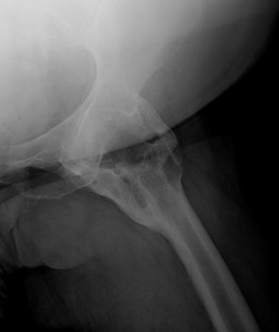
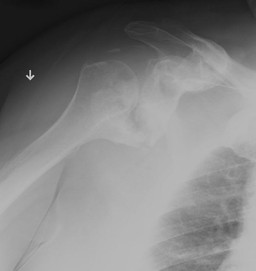
Findings:

BONE SCINTIGRAPHY (LIMITED)

RADIOPHARMACEUTICAL: 20.1 mCi Tc-99m MDP i.v.


FINDINGS: The cervical spine, chest, abdomen, pelvis and proximal thighs were evaluated with delayed images. There is increased radiotracer activity within the right humeral head and under the clavicle. There is increased radiotracer uptake around the left femoral head. There is increased uptake within the bilateral patellofemoral joints consistent with degenerative changes.


LEFT HIP RADIOGRAPH

The left hip demonstrates severe heterotopic ossification with complete bony fusion around the joint.


RIGHT SHOULDER RADIOGRAPH

There is exuberant heterotopic ossification extending from the superior aspect of the right coracoid process fusing with the right clavicle. In addition, there is heterotopic ossification extending from the right glenohumeral joint and the adjacent inferior right scapula extending to and partially fusing with the right humeral head.

Diagnosis: Active heterotopic ossification of the left hip and right shoulder.
General Discussion:

HISTORY

Patient is a 48-year-old woman with history of necrotizing fasciitis of the vulva status post wound debridement with radical vulvectomy and panniculectomy with prolonged ICU stay. Patient has had persistent left hip and right shoulder pain since that time.


DIFFERENTIAL DIAGNOSIS: Localized bone scintigraphy uptake (Mettler, 2006)

trauma

degenerative changes (arthritis)

infection

primary bone tumor (benign and malignant)

metastatic disease

Paget's disease, fibrous dysplasia

hyperemia

overlying soft tissue activity

decreased overlying soft tissue (decreased attenuation)


DISCUSSION: Heterotopic Ossification

Heterotopic ossification is a process in which bone is formed in tissue that does not normally ossify.  Heterotopic ossification is most commonly seen following a neurological disorder or following joint surgery.  Heterotopic ossification has been reported in heavily sedated and immobilized patients in the abscence of a neurological disorder, although it is unusual.  This is the likely etiology in this patient.  There are two proposed pathogenic processes in immobilized patients.  One is that prolonged sedation and immobilization can induce a pathogenic process similar to heterotopic ossification.  The second proposed mechanism is that long term ventilation can cause changes to the local tissue environment, including PO2 and pH, which can then result in heterotopic ossification.  Acute heterotopic ossification can present as pain, fever, swelling, erythema, warmth, stiffness, and/or decreased range of motion.  Heterotopic ossification must be mature prior to treatment/surgical resection in order to minimize postoperative recurrence.  Serial bone scintigraphy can be used to assess the ratio of activity within the heterotopic ossification versus normal bone.  Active heterotopic ossification will demonstrate increased tracer localization on bone scintigraphy.  As the heterotopic ossification matures, the degree of tracer localization will decrease.  A declining ratio suggests maturation.  Follow-up bone scintigraphy is normally performed at 6 months for neurogenic etiology and 18 months for traumatic etiology.

References:

Mettler, Fred and Milton Guiberteau. Essentials of Nuclear Medicine Imaging. 5th ed. Philadelphia, PA: Saunders Elsevier, 2006, pp 244.


Soudry, G and David Drum. Bone Scintigraphy for Evaluation of Heterotopic Ossification in Patients with Spinal Cord Injury, 1993.


Sugita, A., J. Hashimoto, A. Maeda, J. Kobayashi, M. Hirao, K. Masuhara, M. Yoneda, and H. Yoshikawa. Heterotopic Ossification in Bilateral Knee and Hip Joints After Long-Term Sedation. J Bone Miner Metab (2205) 23:329-332.

Comments:
No comments posted.
Additional Details:

Case Number: 271472Owner(s): kwinkl01 and Delphine ChenLast Updated: 02-07-2013
Anatomy: Skeletal System   Pathology: Trauma
Modality: Conventional Radiograph, Nuc MedAccess Level: Readable by all users, writable by NucMed Certifiers
Keywords: bsnm

Case has been viewed 19 times.
Certified by Delphine Chen on 10-06-2010

The reader is fully responsible for confirming the accuracy of this content.
Text and images may be copyrighted by the case author or institution.