Return to Case List with Diagnoses or Case List as Unknowns

FIBROUS DYSPLASIA
Authored By: kwinkl01 and Barry Siegel, Prof of Radiology.
Patient: 67 year old male
History: 67-year-old man with prostate cancer. Evaluate for osseous metastatic disease.
Image Size:[small][as-submitted]

Fig. 1
bone scintigraphy

Fig. 2
computed tomography

Fig. 3
magnetic resonance imaging (T1 post contrast)

Fig. 4
magnetic resonance imaging (T2 post contrast)
Image Size:[small][as-submitted]

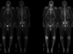
Findings:

BONE SCINTIGRAPHY (WHOLE-BODY)

RADIOPHARMACEUTICAL:  20.1 mCi Tc-99m MDP i.v. 

Delayed whole-body scintigrams were obtained.  


The images demonstrate focally increased activity in the left posterior ilium, appearing as a ring of increased activity with a central area of relatively decreased activity. 


The scintigrams also demonstrate foci of mildly increased activity along the margins of the lower thoracic spine, most likely due to degenerative changes. Increased activity is also seen in the medial compartments of both knees, right greater than left, and in the patellofemoral joints, right greater than left. Small foci of increased activity are seen in the region of the left medial malleolus and the left midfoot. All of the above changes are most likely related to degenerative joint disease. The more intense uptake in the region of the right knee likely reflects the recent right knee arthroscopic surgery. 

There is no definite evidence to suggest osseous metastatic disease.


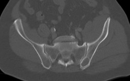
COMPUTED TOMOGRAPHY

Rounded lucent lesion with a well-defined sclerotic margin in the left posterior ilium; the contents of this lesion were of soft tissue density.


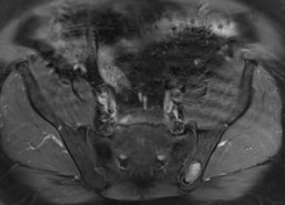
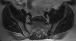
MAGNETIC RESONANCE IMAGING

Rounded lesion within the left posterior ilium.  The lesion is heterogenously hypo- to isointense on T2-weighted imaging and is likely enhancing on post-contrast T1-weighted imaging.  Note that there are no pre-contrast T1 weighted images through this lesion, as the pelvic MRI was performed utilizing a prostate imaging protocol.  Overall findings are consistent with a soft tissue lesion rather than a cystic one.

Diagnosis:

Fibrous dysplasia of the left ilium is the most likely explanation for this finding.


This lesion was stable in appearance by comparison with bone scintigraphy and pelvic CT performed two years ealier when the prostate cancer was first diagnosed.  However, the uptake on bone scintigraphy was incorrectly thought to be related to sacroiliac joint degenerative changes and the lesion on CT was thought to be a degenerative cyst.

General Discussion:

HISTORY:  67-year-old man with prostate cancer, post prostatectomy. He now  has a PSA recurrence (PSA 0.4 mg/dL). Evaluate for osseous metastatic disease.


DIFFERENTIAL DIAGNOSIS: Localized bone scintigraphy uptake (Mettler, 2006)

trauma

degenerative changes (arthritis)

infection

primary bone tumor (benign and malignant)

metastatic disease

fibrous dysplasia, Paget's disease

hyperemia

overlying soft tissue activity

decreased overlying soft tissue (decreased attenuation)


DISCUSSION:  Fibrous dysplasia is an osseous disorder, in which normal bone is replaced with fibrous tissue.  There are no known etiologies.  As the lesion grows, the fibrous tissue causes the bone to expand and deform.  The fibrous tissue also causes the bone to weaken, as the fibrous tissue is softer and not uniform, when compared to the trabecular bone that it replaced.  This causes the bone to be more susceptible to fracture.  Fibrous dyplasia can be monostotic versus polystotic.


Fibrous dysplasia may not cause any signs or symptoms, if mild.  No symptoms or mild symptoms typically occur in patients with monostotic disease.  Most monostotic disease processes are incidentally seen on imaging performed for other reasons.  More severe, polystotic, forms of fibrous dysplasia can present with bone pain, bone deformities, fractures, and difficulty walking.


Conventional radiographs typically show a lytic lesion with an underlying "ground-glass" appearance.  Computed tomography and magnetic resonance imaging can be used to assess the extent of the lesion; degree of bone involvement, associated nerve involement.  Bone scintigraphy is useful in assessing monostotic versus polystotic disease.


Treatment with bisphosphonates can be used to limit osseous breakdown and preserve bone mass.  Surgery maybe performed to correct deformites or fixate fractures.  Surgery can also be performed to remove fibrous tissue, particularly if the fibrous tissue is causing nerve impingement.

References: Mettler, Fred and Milton Guiberteau. Essentials of Nuclear Medicine Imaging. 5th ed. Philadelphia, PA: Saunders Elsevier, 2006, pp 244.
Comments:
No comments posted.
Additional Details:

Case Number: 270977Owner(s): kwinkl01 and Barry Siegel, Prof of RadiologyLast Updated: 02-07-2013
Anatomy: Skeletal System   Pathology: Neoplasm
Modality: CT, MR, Nuc MedAccess Level: Readable by all users, writable by NucMed Certifiers
Keywords: bsnm; 4.355

Case has been viewed 22 times.
Certified by Barry Siegel on 10-10-2010

The reader is fully responsible for confirming the accuracy of this content.
Text and images may be copyrighted by the case author or institution.