Return to Case List with Diagnoses or Case List as Unknowns

RECURRENT OSTEOSARCOMA (BONE SCINTIGRAPHY)
Authored By: Keith Fischer and Andrew Homb.
Patient: 19 year old male
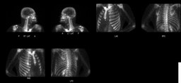
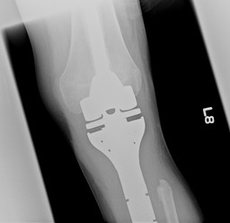
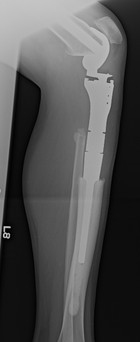
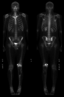
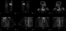
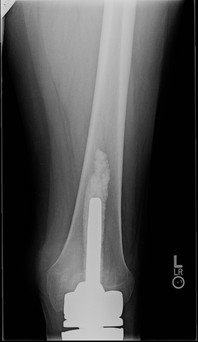
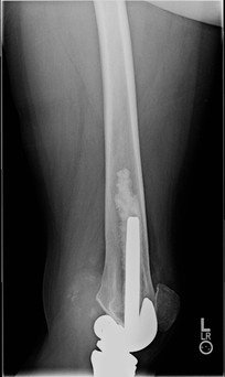
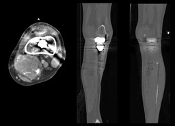
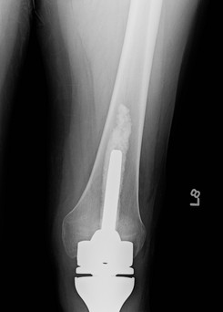
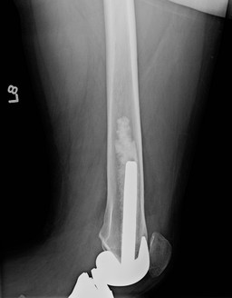
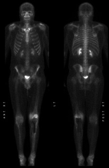
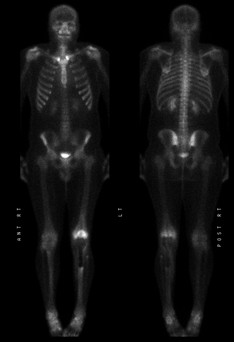
History:

19 year old boy with left tibial osteosarcoma status post excision, prothesis placement, and chemotherapy.  Routine follow-up after completion of therapy.


Nine months after completion of therapy:


Figure 1

Figure 2


Corresponding radiographs


Figure 3

Figure 4


What is your interpretation of this study?


1 year after completion of therapy 


Figure 5

Figure 6


Corresponding radiographs


Figure 7

Figure 8


What is you interpretation of this study and how has it changed since the prior study?


Corresponding CT images (1 year after completion of therapy)


Figure 9


Image Size:[small][as-submitted]

Fig. 1
Whole Body Bone Scintigraphy (9 months after completion of initial therapy)

Fig. 2
Whole Body Bone Scintigraphy (9 months after completion of initial therapy)

Fig. 3
Frontal Radiograph (9 months after completion of initial therapy)

Fig. 4
Lateral Radiograph (9 months after completion of initial therapy)

Fig. 5
Whole Body Bone Scintigraphy (1 year after completion of initial therapy)

Fig. 6
Whole Body Bone Scintigraphy (1 year after completion of initial therapy)

Fig. 7
Frontal Radiograph (1 year after completion of initial therapy)

Fig. 8
Lateral Radiograph (1 year after completion of initial therapy)

Fig. 9
CT images (1 year after completion of initial therapy)

Fig. 10
Frontal Radiograph (3 months after the second resection)

Fig. 11
Lateral Radiograph (3 months after the second resection)

Fig. 12
Whole Body Bone Scintigraphy (3 months after the second resection)

Fig. 13
Whole Body Bone Scintigraphy (7 months after the second resection)
Image Size:[small][as-submitted]

Findings:

Bone Scinitgraphy (9 months after completion of therapy)


RADIOPHARMACEUTICAL: 18.3 mCi Tc-99m MDP i.v.


No evidence of osseous metastatic disease.


Partially visualized new radiotracer uptake within the left fourth and fifth metatarsals which is likely secondary to altered biomechanical stress. If clinically indicated, x-rays of the foot can be obtained for further evaluation.


 Radiographs (9 months after completion of therapy)


No evidence of tumor recurrence status post osteosarcoma resection with modular reconstruction.


Bone Scintigraphy (1 year after completion of therapy)


RADIOPHARMACEUTICAL: 20.2 mCi Tc-99m MDP i.v.


Interval development of abnormally increased radiotracer uptake in the soft tissues of the left knee medial to the distal left femur.


Interval decrease of the previously noted radiotracer uptake in the left foot is consistent with improvement in stress change on that foot from altered weight bearing.


Radiographs (1 year after completion of therapy)


Incompletely seen constrained modular type total left knee arthroplasty. The visualized instrumentation is intact without surrounding lucency or periprosthetic fracture. There is soft tissue prominence with increased ossific densities in the popliteal fossa.


CT Left Lower Extremity (1 year after completion of therapy)


Lobulated soft tissue mass containing calcification posterior to the medial femoral condyle. This mass measures 6.4 x 4.5 cm transaxially and 6.0 cm craniocaudal and is located deep to the semitendinosus tendon and inseparable from the semimembranosus muscle. The mass is separate from the neurovascular bundle and no adjacent bone destruction is seen. There has been excision of the proximal tibia with prosthetic replacement and a constrained left knee arthroplasty.

DDx:

Reccurent osteosarcoma in the soft tissues


Myositis ossificans

Diagnosis: Recurrent Osteosarcoma (Surgical Pathology)
General Discussion:

The patient underwent a second surgical resection for recurrence several weeks after the 1 year post initial treatment bone scintigraphy study. 


Follow up radiographs (3 months after the second resection)


Figure 10

Figure 11


Ressection of the soft tissue prominence with increased ossific densities in the left popliteal fossa.


Bone Scintigraphy (3 months after the second resection)


Figure 12


RADIOPHARMACEUTICAL: 20.8 mCi Tc-99m MDP i.v.


Small amount of residual radiotracer uptake in the soft tissues of the left knee medial to the distal left femur; the low-level of uptake suggests that this is most likely postoperative change and less likely residual tumor. No evidence of distant metastatic disease identified.


Bone Scintigraphy (7 months after the second resection)


Figure 13


RADIOPHARMACEUTICAL: 21.6 mCi Tc-99m MDP i.v.


Radiotracer uptake in the distal femur and proximal fibula, most consistent with postoperative change.


Interval decrease in radiotracer uptake in the soft tissues medial to the distal left femur.


Specific Discussion:

Full History:


19 year old boy with left tibial osteosarcoma status post excision, prothesis placement, and chemotherapy.  Routine follow-up after completion of therapy.  The patient developed recurrence in the soft tissues of the distal left thigh one year after completion of initial therapy and underwent surgical resection.  Follow up studies after the second surgical resection are under the general discussion tab.


Discussion: Recurrent Osteosarcoma


Study by Bacci et al. retrospectively evaluated the pattern of relapse, treatment, and final outcome of 235 patients with non-meatastaic osteosarcoma of the extremity initially treated with neoadjuvant chemotherapy and surgery, who then subsequently relapsed.


In the study: 379 patients did not develop recurrence (61.1%), 6 patients died of chemotherapy toxicity, and 235 patients relapsed.


Pattern of Relapse


Isolated lung metastases: 80%


Isolated distant bone metastases: 8.9%


Isolated local recurrence: 7.6%


Isolated metastasis to another site: 0.8%


Local recurrence with lung metastasis 5.9%


The median interval between start of treatment and the 1st recurrence was 25.4 months


The median interval between the 1st and 2nd recurrence was 12.7 months 


Most patients were treated with surgery (60%) and chemotherapy only as needed.


173 patients achieved remission after treatment (73.6%); however, 120 of these patients relapsed (~70%)

References: Bacci et al. "Treatment and outcome of recurrent osteosarcoma: experience at Rizzoli in 235 patients initially treated with neoadjuvant chemotherapy." Acta Oncol 2005;44(7):748-55.
Comments:
No comments posted.
Additional Details:

Case Number: 245553Owner(s): Keith Fischer and Andrew HombLast Updated: 02-07-2013
Anatomy: Skeletal System   Pathology: Neoplasm
Modality: CT, Nuc MedAccess Level: Readable by all users, writable by NucMed Certifiers
Keywords: bsnmACR: 40000.32210

Case has been viewed 33 times.
Certified by Keith Fischer on 09-03-2010

The reader is fully responsible for confirming the accuracy of this content.
Text and images may be copyrighted by the case author or institution.