Return to Case List with Diagnoses or Case List as Unknowns

BREMSSTRAHLUNG RADIATION INTERFERENCE FROM PREVIOUS Y-90 MICROSPHERES TREATMENT ON A HEPATIC PERFUSION STUDY
Authored By: Andrew Homb and Jerold Wallis, Assoc Prof of Radiology.
Patient: 52 year old male
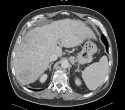
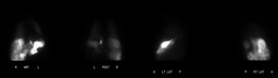
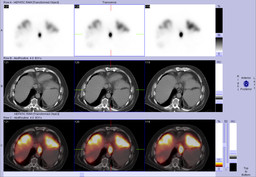
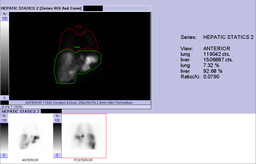
History:

52 year-old man with metastatic colorectal cancer. 


CT


 Figure 1


Impression? 


What therapy can be offered to this patient?


Figure 2

Figure 3

Figure 4


How and why do we perform this study? Interpretation?


Three Weeks later


The patient was treated with Y-90 microspheres to the right hepatic lobe.  


Immediately after Y-90 microsphere treatment another MAA injection was performed into the replaced left hepatic artery to evaluate previously described esophagogastric junction tracer activity, attributed to reflux into the left gastric artery.


Figure 5

Figure 6

Figure 7

Figure 8

Figure 9


Interpretation? 


What artifact is present and what is its cause?


Image Size:[small][as-submitted]

Fig. 1
Axial CT

Fig. 2
Hepatic perfusion scintigraphy (Initial study prior to treatment)

Fig. 3
Hepatic perfusion scintigraphy (Initial study prior to treatment)

Fig. 4
Hepatic perfusion scintigraphy (Initial study prior to treatment)

Fig. 5
Hepatic Perfusion (Three weeks after the initial study)

Multimedia: 245511_6_submitted.avi
MIP cine of SPECT Hepatic Perfusion (Three weeks after the initial study)

Fig. 7
Hepatic Perfusion (Three weeks after the initial study)

Fig. 8
Hepatic Perfusion (Three weeks after the initial study)

Fig. 9
Hepatic Perfusion (Three weeks after the initial study)

Fig. 10
Hepatic perfusion camera setup and Yttrium information (Y-90 and other isotopes)
Image Size:[small][as-submitted]

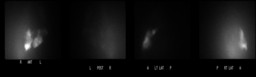
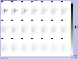
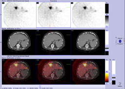
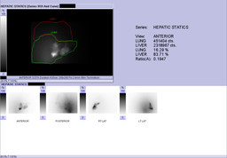
Findings:

Hepatic Perfusion Scinitigraphy (Initial Study prior to therapy)


RADIOPHARMACEUTICAL: 5.3 mCi Tc-99m MAA i.a.


1. Following intra-arterial infusion of Tc-99m MAA particles into the catheterized hepatic artery, there is heterogeneous perfusion of the right and left hepatic lobes. There is relatively higher tracer deposition in the left hepatic lobe. 


2. Intense tracer deposition in the distal esophagus and gastroesophageal junction, indicating extrahepatic perfusion of these structures during Tc-99m MAA injection, likely related to the left replaced hepatic artery injection, with reflux into the left gastric artery.


3. No significant pulmonary activity. The relative lung perfusion is 7% of the injected dose.


Angiographic Y-90 Microsphere Treatment (3 weeks after the initial study)


1. Successful Sirtex radioembolization of the right hepatic artery.


2. Successful repeated injection of MAA to the replace left hepatic artery (to clarify if the previous tracer activity at the level of the gastroesophageal junction was due to reflux to the left gastric artery).  


Hepatic Perfusion Scinitigraphy (3 weeks after the initial study)


RADIOPHARMACEUTICAL: 2.4 mCi Tc-99m MAA i.a. 


1. Following intra-arterial infusion of Tc-99m MAA particles into the catheterized left hepatic artery, there is patchy heterogenous perfusion of the left hepatic lobe.


2. There is significant scatter radiation, thus not allowing the calculation of relative lung perfusion.


3. No discrete tracer deposition was identified in the distal esophagus and gastroesophageal junction on this exam to suggest perfusion of this region via the hepatic artery injection.

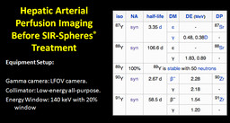
DDx:

Bremsstahlung radiation from concurrent Y-90 administration


Other possible artifacts that might give a similar appearance:


FDG PET patient (unshielded) adjacent to the imaging room


Other recent higher energy imaging study


Diagnosis: Significant scatter radiation secondary to bremsstrahlung radiation originating from Y-90 microspheres injection performed just prior a Tc99m MAA injection and imaging.
General Discussion:

Full History:


52 year-old man with colorectal cancer metastatic to the liver.  The patient is status post surgery and chemotherapy and is now undergoing Y-90 microsphere treatment.  Initial hepatic perfusion scintigraphy demonstrated tracer activity at the gastroesophageal junction indicating extrahepatic perfusion.  Three weeks later the patient received Y-90 microspheres treatment to the right hepatic lobe.  Repeat hepatic perfusion scintigraphy was performed, immediately after Y-90 treatment, with injection of Tc99m MAA into a replaced left hepatic artery to clarify if the previously noted extrahepatic perfusion to the upper GI tract indicated a real (antegrade) perfusion issue, or simply represented backflow into the left gastric artery, possibly related to high flow rates at the time of injection. 


Discussion:


Bremsstrahlung radiation in the context of Y-90 beta emission: broad spectrum secondary gamma-ray emissions produced as a result of the interaction of the high-energy beta-emissions with tissue.


"Beta particle-emitting substances sometimes exhibit a weak radiation with continuous spectrum that is due to Bremsstrahlung. In this context, Bremsstrahlung is a type of "secondary radiation", in that it is produced as a result of stopping (or slowing) the primary radiation (beta particles). In electron and positron emission the photon's energy comes from the electron/nucleon pair, with the spectrum of the bremsstrahlung decreasing continuously with increasing energy of the beta particle. In electron capture the energy comes at the expense of the neutrino, and the spectrum is greatest at about one third of the normal neutrino energy, reaching zero at zero energy and at normal neutrino energy."  Wikipedia (http://wn.wikipedia.org/wiki/Bremsstrahlung)


Images of bremsstrahlung radiation may be obtained using a medium imaging collimator within 1-2 days after Y-90 administration to detect extrahepatic deposition or to determine the distribution of microsphere in the liver; however, this is not typically done at our site. 


As can be seen in this case, bremsstrahlung radiation from Y-90 beta emission can cause scatter as well as septal penetration when concurrently performed with a Tc99m MAA imaging study (using a low energy collimator).


Figure 10: Hepatic Perfusion imaging in preperation for Y-90 microsphere treatment (Tc99m MAA) and information regarding the radiation characteristics for Y-90 microspheres.  


References: Murthy et al.  "Yttrium-90 Microsphere Therapy for Hepatic Malignancy: Devices, Indications, Technical Considerations, and Potential Complications"  2005. Radiographics; 25: S41-S55.
Comments:
No comments posted.
Additional Details:

Case Number: 245511Owner(s): Andrew Homb and Jerold Wallis, Assoc Prof of RadiologyLast Updated: 02-07-2013
Anatomy: Other   Pathology: Iatrogenic
Modality: Nuc MedAccess Level: Readable by all users, writable by NucMed Certifiers
Keywords: hpnm

Case has been viewed 61 times.
Certified by Jerold Wallis on 07-28-2010

The reader is fully responsible for confirming the accuracy of this content.
Text and images may be copyrighted by the case author or institution.