Return to Case List with Diagnoses or Case List as Unknowns

DIEULAFOY LESION PRESENTING AS GI BLEEDING
Authored By: Andrew Homb and Jerold Wallis, Assoc Prof of Radiology.
Patient: 53 year old female
History:

53 year old female with ischemic cardiomyopathy necesitating placement of a 2nd-generation left ventricular assist device (LVAD). The patient presents with a melena while on warfarin and clopidogrel.  See figures 1-3


What is your interpretation of this study and what is the next step in management?


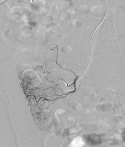
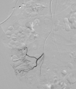
Findings and intervention performed?  (figures 4-9)


What additional procedure may be helpful in determining a diagnosis? (see figure 10)


Image Size:[small][as-submitted]

Multimedia: 245472_1_submitted.avi
Anterior GI Bleeding Scintigraphy Cine

Multimedia: 245472_2_submitted.avi
Posterior GI Bleeding Scintigraphy Cine

Fig. 3
GI Bleeding Scintigraphy

Fig. 4
Pretherapy Angiography 1

Fig. 5
Pretherapy Angiography 2

Fig. 6
Pretherapy Angiography 3

Fig. 7
Therapy Angiography 4

Fig. 8
Therapy Angiography 5

Fig. 9
Post Therapy Angiography 6

Fig. 10
Colonoscopy
Image Size:[small][as-submitted]

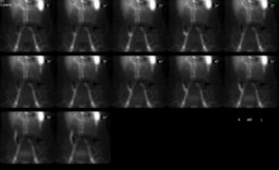
Findings:

Gastro-intestinal Bleeding Scintigraphy:

RADIOPHARMACEUTICAL: 26.8 mCi Tc-99m in vitro labeled red cells i.v.

Lower gastrointestinal hemorrhage, specifically arising from the region of the cecum.


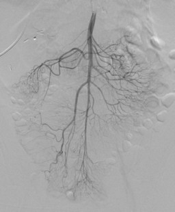
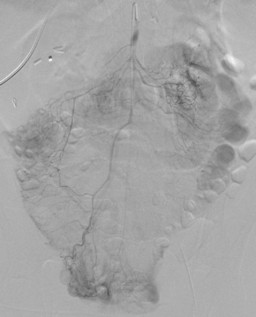
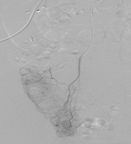
Angiography:

Coil embolization of distal branch of the ileocolic artery supplying the bleeding segment of the cecum. No evidence of active bleeding on the post coiling angiograms.


Colonoscopy:

See figure 10 and general discussion

DDx:

Dieulafoy lesion


Angiodysplasia


Other causes of lower gastrointestinal bleeding:


Diverticular disease


Telangectasias


Neoplasms


Advanced liver disease causing mucosal spider nevi


Diagnosis: Dieulafoy Lesion
General Discussion:

Full History:

53 year old female with ischemic cardiomyopathy necesitating placement of a 2nd-generation left ventricular assist device (LVAD). The patient presented with a G.I. bleed while on warfarin and clopidogrel.  The initial GI bleeding study was positive but the initial angiogram and colonscopy were negative (images not included in this teaching file).  The patient was subsequnetly sent home off of warfarin and clopidogrel but returned with a falling hematocrit and melena.  Both the repeat GI bleeding study and angiography study were positive (images included in this teaching file).  Repeat colonscopy was performed after the angiographic intervention demonstrating the Dieulafoy lesion, which was then treated endoscopically with epinephrine and clipping.

Discussion: Dieulafoy “ulcers”

Large  tortuous arteriole in the submucosa that erodes the gastrointestinal wall. 

< 5% of causes for gastrointestinal bleeding

Location

  • 75% occur along the lesser curvature of the upper part of the stomach
  • 14% duodenum,
  • 5% colon,
  • 5% surgical anastamoses

No direct association alcohol abuse or NSAID use.

Treatment is primarily by endoscopic techniques

References: Jain, Richa and Chetty, Runjan. "Dieulafoy Disease of the Colon." Arch Pathol Lab Med. 133; 1865-1867, 2008.
Comments:
No comments posted.
Additional Details:

Case Number: 245472Owner(s): Andrew Homb and Jerold Wallis, Assoc Prof of RadiologyLast Updated: 02-07-2013
Anatomy: Gastrointestinal (GI)   Pathology: Vascular
Modality: Nuc Med, AngiographyAccess Level: Readable by all users, writable by NucMed Certifiers
Keywords: ginm

Case has been viewed 44 times.
Certified by Jerold Wallis on 07-28-2010

The reader is fully responsible for confirming the accuracy of this content.
Text and images may be copyrighted by the case author or institution.