General Discussion: Full Patient History:
Patient is a 55 year-old man with a diagnosis of appendicular mucinous adenocarcinoma with associated pseudomyxoma peritonei. He is status post total proctocolectomy with end ileostomy, small bowel resection, debulking of carcinomatosis, and omentectomy. Two intraperitoneal catheters were placed in anticipation of intraperitoneal chemotherapy.
General Discussion:
Peritoneal cavity scintigraphy is indicated for (1) assessing whether there is direct communication between the peritoneal cavity and an extraperitoneal fluid collection (e.g., pleural effusion , hydrocele) and (2) evaluating the intraperitoneal distribution of tracer before intracavitary therapy of malignant ascites with intraperitoneal chemotherapy (P-32 chromic phosphate colloid intraperitoneal radiotherapy is seldom performed now). Typically the radiopharmaceutical used is Tc-99m-sulfur colloid.
A particulate radiopharmaceutical injected into the peritoneal cavity will normally distribute throughout this space. Abnormal connections of the peritoneal cavity with the pleural space or the scrotum are demonstrated by the presence of tracer in these regions after peritoneal injection.
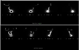
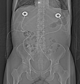
For patients who had prior abdominal surgery, as in this case, peritoneal cavity scintigraphy is particularly important, since subsequent adhesions might prevent flow of peritoneal fluid, decreasing the effectiveness of intraperitoneal chemotherapy. To best evaluate fluid flow, the patient is asked to lie in decubitus positions, and to walk around for several minutes after radiotracer injection. Free radiotracer movement in the peritoneal cavity might not produce an outline of the cavity, but activity should not be seen just around the catheter tip. In this case, loculated fluid is seen in the lower abdomen, next to the catheter tips, after injections into both left and right catheters. The position of the catheter tip was confirmed on radiograph. No free-flow of fluid is seen. Thus, based on this study, intraperitoneal chemotherapy was not initiated. Patient continued to receive systemic chemotherapy.