Return to Case List with Diagnoses or Case List as Unknowns

REDUCED CEREBRAL PERFUSION AFTER BALLOON OCCLUSION OF CAROTID ARTERY
Authored By: Collin Liu and Jerold Wallis, Assoc Prof of Radiology.
Patient: 61 year old male
History: 61-year-old man with left squamous cell tonsillar carcinoma status post radical pharyngectomy and base of tongue resection with known invasion of the left carotid artery.
Image Size:[small][as-submitted]

Fig. 1
Brain SPECT Day #1 (injection during balloon occlusion)

Fig. 2
Brain SPECT Day #1 (injection during balloon occlusion)

Fig. 3
Brain SPECT Day #2 (baseline exam)

Fig. 4
Brain SPECT Day #2 (baseline exam)
Image Size:[small][as-submitted]

Findings:

Brain SPECT #1:

Radiopharmceutical: 27 mCi Tc-99m bicisate via peripheral i.v., during left carotid occlusion 

Diffuse, 10 to 15% reduction in perfusion of the left frontal and parietal lobes following left carotid artery balloon occlusion, with smaller more focal areas of 20% reduction in perfusion in the left frontal lobe when compared to the contraleral side (Figure 1 and Figure 2).


Brain SPECT #2:

Radiopharmceutical: 25 mCi Tc-99m bicisate i.v.

There is a symmetrical FDG uptake in the cortices. The prior areas of decreased perfusion in the left frontal and parietal lobes seen on the brain SPECT #1 are not visualized on today's study (Figure 3 and Figure 4).

Diagnosis: 10-20% drop in cerebral perfusion after temporary balloon occlusion
General Discussion:

Full Patient History:
61-year-old man with left squamous cell tonsillar carcinoma status post radical pharyngectomy and base of tongue resection with known invasion of the left carotid artery. Radiation therapy has a risk of left carotid artery injury. Therefore, left carotid artery balloon occlusion was performed to evaluate for significant drop in cerebral perfusion

General Discussion:
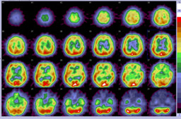
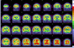
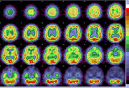
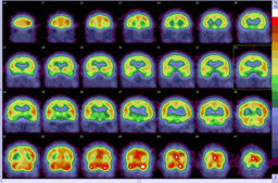
Temporary balloon-occlusion SPECT using Tc-99m bicisate or Tc-99m HMPAO allows evaluation of cerebral vascular reserve for potential carotid artery sacrifice. 

Normal cerebral blood flow (CBF) is approximately 55 ml/100 g/min. Neuronal dysfunction and cerebral ischemia develop when CBF drops below about 20 ml/100 g/min with cerebral infarction occurring when CBF falls below 10 ml/100 g/min. The appearance of a  neurologic deficit during balloon occlusion of the ICA, with simultaneous neurologic examination, indicates that CBF has fallen below the critical threshold necessary to maintain neuronal function and is considered unequivocal evidence that the patient will not tolerate ICA occlusion. Unfortunately the failure to develop neurologic defects during temporary balloon occlusion does not ensure the safety of the procedure and stroke rates of up to 20% following a negative temporary balloon occlusion have been reported. Thus Tc-99m bicisate or Tc-99m HMPAO SPECT is useful in assessing significant drop in perfusion in asymptomatic patients.

In this case, the patient did not demonstrate any decline in his neurological status during 30 minutes of occlusion. The occlusion study (SPECT Day #1) demonstrated 10-20% drop in cerebral perfusion in the left hemisphere, compared to the right. The baseline study (SPECT Day #2) performed the next day showed symmetrical perfusion. This suggests that the perfusion asymmetry seen on the occlusion study was due to carotid artery occlusion. However, because of limited treatment alternatives, the patient subsequently received complete embolization of the cervical segment of left internal carotid artery. Before discharge, he developed transient right sided facial drop and weakness with low blood pressure, indicating low cerebral vascular reserve in the left hemisphere. The patient was discharged on anticoagulation therapy.


References:

Lorberboym M, Pandit N, Machac J. et al. The role of brain perfusion imaging during preoperative temporary balloon occlusion of the internal carotid artery. J Nuc Med
1996:37:415-419.

Origitano TC, Al-Mefty O, Leonetti JP, DeMonte F, Reichman OH. Vascular considerations and complica tions in cranial base surgery. Neurosurgery 1994;35: 351-363.

Comments:
No comments posted.
Additional Details:

Case Number: 241833Owner(s): Collin Liu and Jerold Wallis, Assoc Prof of RadiologyLast Updated: 02-07-2013
Anatomy: Cranium and Contents   Pathology: Iatrogenic
Modality: MR, Nuc Med, 3D ReconstructionAccess Level: Readable by all users, writable by NucMed Certifiers
Keywords: cerebral perfusion, balloon occlusion, pbnm, tumor invasion of carotid artery

Case has been viewed 21 times.
Certified by Jerold Wallis on 07-28-2010

The reader is fully responsible for confirming the accuracy of this content.
Text and images may be copyrighted by the case author or institution.