Return to Case List with Diagnoses or Case List as Unknowns

METASTATIC THYROID CANCER
Authored By: tdeshm01 and Henry Royal.
Patient: 39 year old female
History: 39 year old female: status post thyroidectomy for multinodular goiter 8 years ago.  Pathology negative for malignancy.
Image Size:[small][as-submitted]

Fig. 1
WHOLE-BODY I-131 IMAGING

Fig. 2
WHOLE-BODY I-131 IMAGING

Fig. 3
SPECT-CT

Fig. 4
SPECT-CT

Fig. 5
SPECT-CT

Fig. 6
SPECT-CT
Image Size:[small][as-submitted]

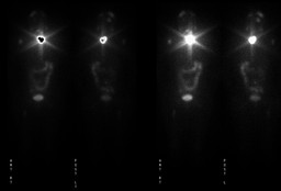
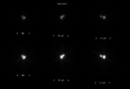
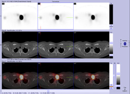
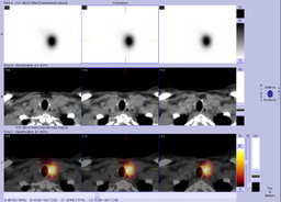
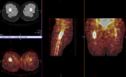
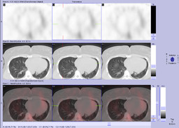
Findings:

RADIOPHARMACEUTICAL: 5.3 mCi I-131 sodium iodide p.o.


1. Most intense I-131 activity in the left neck that likely represents residual thyroid tissue.


2. Increased I-131 activity in the right humeral head, and bilateral proximal femurs, more intense and larger on the right, consistent with osseous metastatic disease.


3. Increased I-131 activity in both sides of the head which may represent scalp or calvarial metastatic disease.


4. Multiple indeterminate bilateral pulmonary nodules, largest in the left lower lobe.

Diagnosis: Metastatic well-differentiated papillary thyroid carcinoma (proven by surgical excision of scalp mass)
General Discussion:  6 years after thyroidectomy for multinodular goiter, the patient noticed an occipital mass that was biopsied and negative for malignancy. She noted that the mass was growing in size over one year.  Surgical excision of the mass was performed and pathology confirmed metastatic thyroid cancer. She was treated with 200 mCi radioactive iodine.
Comments:
No comments posted.
Additional Details:

Case Number: 241186Owner(s): tdeshm01 and Henry RoyalLast Updated: 02-07-2013
Anatomy: Face and Neck   Pathology: Neoplasm
Modality: Nuc MedAccess Level: Readable by all users, writable by NucMed Certifiers
Keywords: trnm

Case has been viewed 24 times.
Certified by Henry Royal on 10-10-2010

The reader is fully responsible for confirming the accuracy of this content.
Text and images may be copyrighted by the case author or institution.