|
| Patient: 8 year old female |
| History: 8-year-old girl with left arm pain for a few weeks. |
Image Size:
|
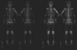
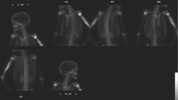
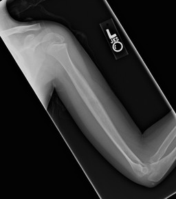
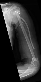
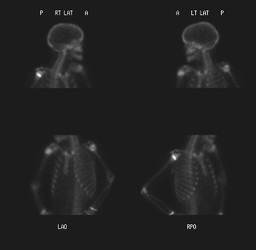
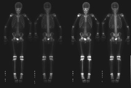
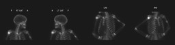
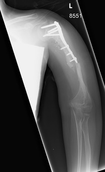
| Findings: Bone scintigraphy: There is marked diffuse increased uptake throughout the humeral diaphysis which appears moderately expanded. Differential diagnosis includes a primary neoplasm and osteomyelitis. Ā ĀRadiograph of left humerus:ĀThere is a permeative, lytic lesion involving the diaphysis of the left humerus with associated aggessive periosteal reaction and a wide zone of transition. Differential considerations include Ewing's sarcoma, PNET, lymphoma, and somewhat less likely osteomyelitis or Langerhans cell histiocytosis. Ā Surgical pathology: Ewing's sarcoma Ā IMAGING POST-SURGERY: Ā Radiograph of left arm: Resection of the humerus from the proximal to distal metaphyses. A fibular bone graft spans this gap. Numerous surgical staples and a surgical drain are in place. Ā Bone scintigraphy (One year post surgery and chemotherapy): Stress reactions within the tibia bilaterally likely secondary to prior right fibular graft harvesting. No evidence of recurrent tumor or metastases. Ā Follow up bone scintigraphy - 9 months after prior exam: Focal increased uptake involving the midportion of the fibular graft in the left upper arm. |
| DDx: Ā 1. FractureĀthrough fibular graft Ā 2. Recurrent tumor |
| Diagnosis: Fracture through the mid-portion of the left arm fibular graft. |
| Comments: No comments posted. |
| Additional Details:
Case Number: 233930 The reader is fully responsible for confirming the accuracy of this content. |