Return to Case List with Diagnoses or Case List as Unknowns

RENAL LACERATION
Authored By: tdeshm01 and Bennett Greenspan.
Patient: 10 year old male
History: 10 year old boy with abdominal trauma.
Image Size:[small][as-submitted]

Fig. 1
Renal Scan

Fig. 2
Renal SPECT:Coronal image

Fig. 3
Renal SPECT:Axial image

Fig. 4
Renal SPECT: Axial image

Fig. 5
CECT Abdomen

Fig. 6
CECT Abdomen
Image Size:[small][as-submitted]

Findings:

RADIOPHARMACEUTICAL: 2.1 mCi Tc-99m DMSA i.v.


FINDINGS: Renal cortical scintigraphy with SPECT was performed. There is a linear photopenic area in the cortex of the lower pole of the left kidney, extending into the interpolar region.The remaining left kidney and right kidney appear well perfused. No injury to the renal artery or renal vein is apparent.

DDx:

Renal Infarct.


Renal Laceration.

Diagnosis: Left renal laceration.
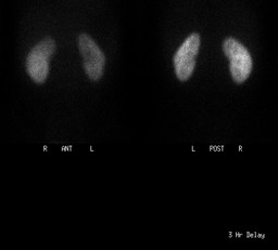
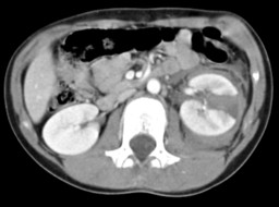
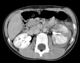
General Discussion:

FULL PATIENT HISTORY:10 year old boy hit in the left flank while playing baseball followed by pain in the left flank and vomiting. CT scan showed grade 4 left renal laceration with no contrast extravasation on delayed images from ureter or bladder. He was managed conservatively, followed up with USG and a renal scan was obtained after 3 months to evaluate renal function. 


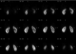
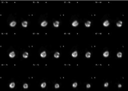
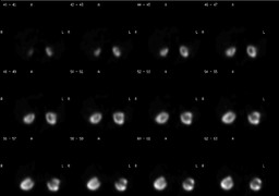
DISCUSSION:Renal cortical imaging is performed by using Tc99m-DMSA or glucoheptonate and a pinhole or high-resolution collimator or SPECT (Single photon emission computed tomography). A pinhole or high-resolution collimator provides very high resolution images. SPECT improves image contrast because it focuses on a thin slice of an organ and minimizes overlying and underlying activity that may obscure the area of interest.


Indications for renal cortical imaging include evaluation of mass lesions, functioning pseudotumors such as cortical columns of Bertin, edema and scarring secondary to pyelonephritis (acute or chronic) especially in children. Follow up of a renal laceration is an uncommon indication of renal cortical scintigraphy; renal functional imaging by using Tc99m-MAG3 or Tc99m-DTPA is preferred.


References: Essentials of Nuclear Medicine Imaging, 5th Edition by Fred A.Mettler,JR. and Milton J. Guiberteau
Comments:
No comments posted.
Additional Details:

Case Number: 198862Owner(s): tdeshm01 and Bennett GreenspanLast Updated: 02-07-2013
Anatomy: Genitourinary (GU)   Pathology: Trauma
Modality: CT, Nuc MedAccess Level: Readable by all users, writable by NucMed Certifiers
Keywords: rsnm

Case has been viewed 33 times.
Certified by Bennett Greenspan on 01-02-2010

The reader is fully responsible for confirming the accuracy of this content.
Text and images may be copyrighted by the case author or institution.