Return to Case List with Diagnoses or Case List as Unknowns

CONGENITAL BILATERAL MEGAURETERS
Authored By: Joanna Fair and Henry Royal.
Patient: 2 month 19 day old
History:

2 month 19 day old patient:

Presenting for urinary tract evaluation.

Image Size:[small][as-submitted]

Fig. 1
Angiographic images from renal scintigraphy.

Fig. 2
Pre-furosemide functional images from renal scintigraphy.

Fig. 3
Post-furosemide functional images from renal scintigraphy.

Fig. 4
Postvoid images (pre- and post- furosemide).

Fig. 5
Renal washin curves.

Fig. 6
Post-furosemide washout curves.

Fig. 7
Bladder image from a voiding cystourethrogram.

Fig. 8
Voiding image from a voiding cystourethrogram.
Image Size:[small][as-submitted]

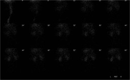
Findings:

(Fig. 1) The posterior abdominal radionuclide angiogram demonstrates normal, symmetrical perfusion of the kidneys.

[pagebreak]


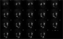
(Fig. 2) Sequential renal images show normal size and morphology of the kidneys, with prompt uptake and excretion bilaterally.  There is mild-to-moderate retained activity in the both collecting systems, which appear of normal to possibly slightly enlarged size.

Both ureters are dilated and tortuous.


[pagebreak]


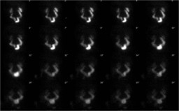
(Fig. 3) To evaluate for obstruction, the patient was given 6 mg furosemide via slow intravenous injection approximately 30 minutes after the start of the examination. Sequential images demonstrate prompt clearance of pelvicalyceal activity bilaterally after diuretic administration.


[pagebreak]


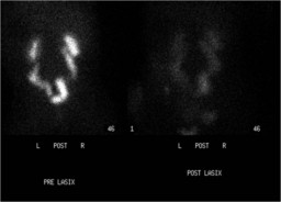
(Fig. 4) The post-furosemide, post-void image shows no significant retained activity.

[pagebreak]


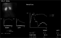
(Fig. 5) The split renal function is nearly symmetric.

[pagebreak]


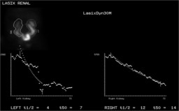
(Fig. 6) After diuretic administration, the half-time of tracer clearance from both kidneys is normal.


[pagebreak]


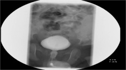
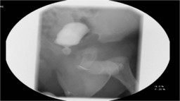
(Fig. 7 and Fig. 8) Voiding cystourethrogram was subsequently performed. This study demonstrated no evidence of vesicoureteral reflux, and no evidence of posterior urethral valves.

DDx:

1. Congenital megaureters.
2. Antenatal obstruction, now relieved.

Diagnosis: Congenital megaureters.
General Discussion:

Full patient history: 

2-month-old boy presenting with hydroureter and hydronephrosis seen on prenatal ultrasound.  Renal scintigraphy was performed to evaluate for obstruction, and the voiding cystourethrogram was performed to evaluated for obstruction.  Based on these images, the etiology of the dilated, nonobstructed ureters was thought to represent congenital megaureter or antenatal obstruction, now relieved.


Discussion: 

A megaureter is a dilated ureter, which can classified be as obstructed, refluxing, obstructed and refluxing, or nonobstructed and nonrefluxing.  Nonrefluxing/nonobstructed megaureter is only diagnosed when reflux and obstruction have been excluded, as in this patient with no evidence of obstruction on renal scintigraphy and no evidence of reflux on voiding cystourethrogram.  Megaureters are typically left-sided when unilateral, are more commonly seen in boys, and are bilateral in approximately 20% of cases. 

References: Merlini E et al, "Primary non-refluxing megaureters," J Pediatr Urol. 2005 Dec;1(6):409-17.

Minevich E and Tackett L, "Megaureter and other congenital ureteral anomalies," www.emedicine.com (updated Feb 6, 2007).
Comments:
No comments posted.
Additional Details:

Case Number: 177199Owner(s): Joanna Fair and Henry RoyalLast Updated: 02-07-2013
Anatomy: Genitourinary (GU)   Pathology: Congenital
Modality: Nuc Med, GUAccess Level: Readable by all users, writable by NucMed Certifiers
Keywords: rsnm, megaureter, megaloureters, renogram, renal scintigraphyACR: 80000.84900

Case has been viewed 31 times.
Certified by Henry Royal on 10-15-2010

The reader is fully responsible for confirming the accuracy of this content.
Text and images may be copyrighted by the case author or institution.