Return to Case List with Diagnoses or Case List as Unknowns

CHOLEDOCHAL CYST
Authored By: Akash Sharma and Jonathan Weiss.
Patient: 8 day old female
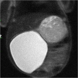
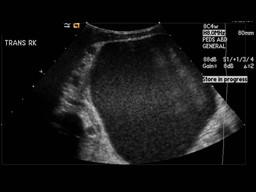
History:

Patient presented with a large abdominal mass.

Image Size:[small][as-submitted]

Fig. 1

Fig. 2
4 hour delayed image

Fig. 3
24 hour delayed image

Fig. 4
T2 weighted coronal MR image

Fig. 5
Ultrasound of the right abdomen (transverse imgae)
Image Size:[small][as-submitted]

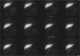
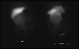
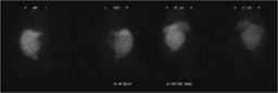
Findings:

Following intravenous administration of 0.6 mCi of Tc-99m mebrofenin, sequential abdominal images were obtained through 60 minutes. There is prompt, uniform accumulation of the tracer by the liver. Images taken through 60 minutes demonstrate the appearance of radiotracer slowly filling the depend portion of the large photopenic mass in the right abdomen (Figure 1).  The gall bladder is not visualized. Delayed images taken at 4 hours demonstrate accumulation of the radiotracer within the right abdomen in a well circumscribed mass which on the early images appeared photopenic (Figure 2).  Repeat images were obtained at 24 hours and demonstrate continued accumulation of radiotracer within the mass (Figure 3).

DDx: Choledochal Cyst
Diagnosis: Choledochal Cyst
General Discussion:

The patient presented with a large cystic mass in the right abdomen.  An MRI and US preformed prior to the nuclear medicine study demonstrates the large cystic mass (Figures 4 and 5).  In order to determine the origin of the mass hepatobiliary imaging was preformed.   The study confirmed that the mass directly communicated with the biliary system.   The mass was a large Type I choledochal cyst (diffuse dilation of the common bile duct).  The large choledochal cyst caused a partial obstruction of the distal common bile duct that was manifested on the haptobiliary scan as absence of radiotracer excrection into the bowel by 24 hours.  The choledochal cyst was subsequently surgically removed.

Comments:
No comments posted.
Additional Details:

Case Number: 172587Owner(s): Akash Sharma and Jonathan WeissLast Updated: 02-07-2013
Anatomy: Gastrointestinal (GI)   Pathology: Congenital
Modality: MR, Nuc MedAccess Level: Readable by all users, writable by NucMed Certifiers
Keywords: hsnm, choledochal cystACR: 00007.14920

Case has been viewed 50 times.
Certified by Akash Sharma on 03-10-2010

The reader is fully responsible for confirming the accuracy of this content.
Text and images may be copyrighted by the case author or institution.