Return to Case List with Diagnoses or Case List as Unknowns

PRIMARY MEGAURETER
Authored By: Henry Royal and Jonathan Weiss.
Patient: 2 year old
Image Size:[small][as-submitted]

Fig. 1

Fig. 2

Fig. 3
After diuretic administration

Fig. 4

Fig. 5
Ultrasound demonstrating the left hydronephrosis and left hydroureter.
Image Size:[small][as-submitted]

Findings:

The posterior abdominal radionuclide angiogram demonstrates normal, symmetrical perfusion of the kidneys (Figure 1).  On sequential renal images, the left kidney is slightly enlarged as compared to the right and the left kidney shows a central area of decreased activity that fills in with radiotracer over time that likely represents hydronephrosis.  There is prompt uptake and excretion of the radiopharmaceutical by the right kidney. The left kidney shows prompt uptake of the radiotracer without significant excretion (Figure 2). 

Prior to diuretic administration, there is moderate retained activity in the left collecting system, which appears moderately enlarged. There is only minimal retained activity in the right collecting system, which appears of normal size.  The left ureter appears markedly dilated and tortuous (Figure 2).

There is prompt clearance of pelvicalyceal activity on the left after diuretic administration. On the right, there is prompt clearance of activity from the pelvicalyceal system (Figure 3).   After diuretic administration, the half-time of tracer clearance from the left kidney is 9 minutes.  The half time of clearance from the right kidney was not calculated as there was near complete tracer clearance from the right kidney even before diuretic administration (Figure 4).


DDx:

Left nonobstructed primary megaureter

Left vesicoureteral reflux

Dilated left collecting system from prior obstruction that has now resolved

Diagnosis:

The patient had recent voiding cystourethrography that did not show reflux; therefore, the most likely diagnosis is a nonobstructed left primary megaureter.

General Discussion:

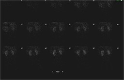
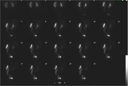
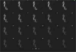
The diuretic renogram is a useful test in determining whether a dilated collection system is functionally obstructed.   Typically the diuretic is administered approximately 20 minutes after radiopharmaceutical administration (typically Tc-99m labeled MAG-3 or DTPA) as long as there is adequate radiotracer within the collecting system in question.   Sequential images are then obtained for an additional 30 minutes.  These images are visually and electronically analyzed to evaluate the clearance of the tracer from the collecting system.

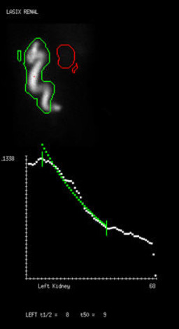
The images are used to calculate a diuretic half-time (T1/2) of the collecting system which is defined as the time at which the time activity curve is decreased to half its original value.  If there is prompted clearance of tracer from the collecting system (T1/2 <10) then there is no functional obstruction.   If the T1/2 is between 10 and 20 minutes, the study is indeterminate for obstruction.   If T1/2 is greater the 20 minutes, the system is likely obstructed.  Visual inspection of the images is also required and should be correlated with the calculated T1/2 and any discrepancies should be resolved.        

The size of the dilated collecting system and renal function must also be taken into account in interpretation.  Decreased renal function and large compliant collecting systems can result in falsely elevated T1/2. 

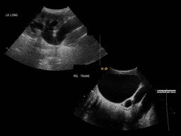
The images can also be used to help determine the location of the obstruction.  As in this case, the left ureter and the colleting system are dilated indicating a possible distal obstruction.  The patient subsequently had a voiding cystourethrogram that showed no reflux.  Therefore, the most likely diagnosis in this patient is a left nonobsructed primary megaureter.


Comments:
No comments posted.
Additional Details:

Case Number: 165159Owner(s): Henry Royal and Jonathan WeissLast Updated: 02-07-2013
Anatomy: Genitourinary (GU)   Pathology: Congenital
Modality: CT, GUAccess Level: Readable by all users, writable by NucMed Certifiers
Keywords: megaloureterACR: 80000.84900

Case has been viewed 22 times.
Certified by Henry Royal on 09-17-2009

The reader is fully responsible for confirming the accuracy of this content.
Text and images may be copyrighted by the case author or institution.