|
| ||||||
|
| ||||||
|
|
|
| Patient: 70 year old |
| History: 70 year old patient: Additional history withheld. |
Image Size:
|
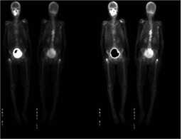
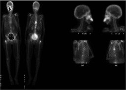
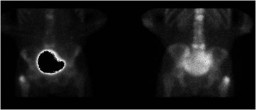
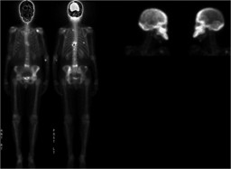
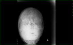
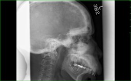
| Findings: Radiopharmaceutical:Ā 19.8 mCi Tc-99m MDP i.v. Whole-body images, spot views, and post-void imagesĀwere obtained. (Fig. 1, Fig. 2, and Fig. 3). These images demonstrate intense calvarial and facial bone uptake. There are also multifocal areas of abnormal uptake in the thoracic and lumbar spine, the left ilium/sacrum,Ābilateral scapulae, the right greater trochanter, and at least one right posterior rib. [pagebreak] A bone scan obtained 1.5 years previously (Fig. 4) demonstrated even more intense calvarial and facial bone uptake, with multifocal abnormal uptake elsewhere in the skeleton. What additional imaging would you like to see? [pagebreak] Frontal and lateral skull radiographs demonstrate a 'cotton-wool' appearance to the calvarium with expansion of the mandible, right greater than left. |
| DDx: Multifocal metastatic disease versus Paget's disease plus multifocal metastatic disease. |
| Diagnosis: Paget's disease plus multifocal metastatic disease from breast cancer. |
| References: Ryan, PJ and Fogelman, I.Ā "Bone Scintigraphy in Metabolic Bone Disease."Ā Semin Nucl Med. 1997 Jul;27(3):291-305. |
| Comments: No comments posted. |
| Additional Details:
Case Number: 148550 The reader is fully responsible for confirming the accuracy of this content. |