Return to Case List with Diagnoses or Case List as Unknowns

PENILE CHLOROMA
Authored By: Joanna Fair and Bennett Greenspan.
Patient: 38 year old
History: Acute myelogenous leukemia; stem cell transplant 14 months previously.
Image Size:[small][as-submitted]

Multimedia: 144871_1_submitted.avi
Whole-body FDG-PET/CT scan

Fig. 2
Images from a whole-body FDG-PET/CT scan

Fig. 3
Images from a whole-body FDG-PET/CT scan

Fig. 4
Images from a whole-body FDG-PET/CT scan

Fig. 5
Images from an MR examination of the pelvis, including pre-contrast T1, T2, and post-contrast T1 sequences.

Fig. 6
Images from an ultrasound of the left biceps muscle long head and left infraspinatus muscle.
Image Size:[small][as-submitted]

Findings: FDG-PET images demonstrate markedly increased FDG uptake in the corpora cavernosa.  There is also increased FDG uptake in the left infraspinatus muscle and left biceps muscle long head.

[pagebreak]

MR images demonstrate an enhancing mass centered at the junction of the extrapenile soft tissues and the corpora spongiosa. There is dilatation of the ureter proximal to this mass.  The mass and the dilated ureter produce the FDG uptake seen in this region on the PET-CT examination.

[pagebreak]

The ultrasound images show no abnormality in the left infraspinatus or in the left biceps muscle long head.

DDx: Extramedullary myeloid tumor (chloroma) versus primary penile malignancy.
Diagnosis: Extramedullary myeloid tumor (chloroma, granulocytic sarcoma)
General Discussion: Full patient history:

38-year-old man with acute myelogenous leukemia (AML).  His disease had been in remission after a matched unrelated donor stem cell transplant 14 months previously.  The patient presented with a palpable penile mass.  The patient did not have any left shoulder pain.  After the MRI, the penile mass was biopsied and found to be an extramedullary myeloid tumor.  Given the lack of symptoms in the left shoulder and the normal ultrasound examination, no biopsy was performed of the left shoulder muscles.

Discussion:
Chloroma (also known as granulocytic sarcoma) is a localized extramedullary
tumor of myeloid cells.  This tumor is most frequently seen as an extramedullary relapse in patients with myelogenous leukemia, with significantly increased risk in patients who have undergone allogeneic stem cell transplantation.  Chloromas can be soft-tissue nodules or diffusely infiltrative masses, and they are frequently found in the breasts, subcutaneous tissues, and bone, although they can be found virtually anywhere.  Imaging features are often similar to lymphoma.  These focal recurrences are typically treated with localized therapy (e.g., radiation). 
References: Fritz, J et al. Am J Roentgenol. 2007 Jul;189(1):209-18.
Comments:
No comments posted.
Additional Details:

Case Number: 144871Owner(s): Joanna Fair and Bennett GreenspanLast Updated: 02-07-2013
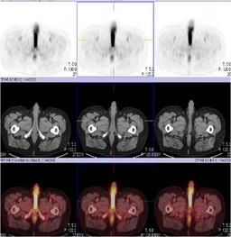
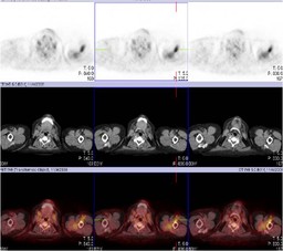
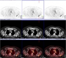
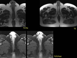
Anatomy: Genitourinary (GU)   Pathology: Neoplasm
Modality: MR, Nuc Med, USAccess Level: Readable by all users, writable by NucMed Certifiers
Keywords: ptnm, acute myelogenous leukemia, aml, chloroma, extramedullary myeloid sarcomaACR: 80000.34100

Case has been viewed 44 times.
Certified by Bennett Greenspan on 06-23-2009

The reader is fully responsible for confirming the accuracy of this content.
Text and images may be copyrighted by the case author or institution.