Return to Case List with Diagnoses or Case List as Unknowns

PLANTAR FASCIITIS
Authored By: Farrokh Dehdashti and Jonathan Weiss.
Patient: 30 year old female
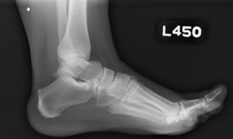
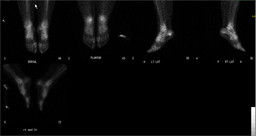
History:

30 year old female:

Chronic left heel pain.

Image Size:[small][as-submitted]

Fig. 1

Fig. 2
Image Size:[small][as-submitted]

Findings:

RADIOPHARMACEUTICAL: 21.6 mCi Tc-99m MDP i.v.

FINDINGS:  The lateral foot radiograph is normal.  On delayed bone scan images of the feet and ankles, there is a small focus of increased radiotracer uptake at the inferior aspect of left calcaneous at the expected origin of the plantar fascia. 

DDx: Plantar fasciitis. 
Diagnosis: Plantar fasciitis.
General Discussion:

Plantar fasciitis is the most common cause of heel pain, reportedly accounting for 11%–15% of all patients with foot pain who present for medical care. The incidence peaks between the ages of 40 and 60 y in the general population and earlier in runners, and approximately one third of cases are bilateral. In most cases, a bone scan is obtained to confirm the diagnosis and to guide the injection of corticosteroids or local anesthetics. Our results confirmed an additional predictive role for the bone scintigram. Focal calcaneal hyperemia is clearly associated with a high success rate for injection, with extension of hyperemia into the proximal soft tissues being associated with a response in only 50% and diffuse hyperemia with no response.

Focal increased radiotracer uptake at the insertion of the plantar fascia is the typical finding in plantar fasciitis.  Radionuclide angiographic and immediate blood pool images can be used to differentiate plantar fasciitis into two types (Type I and Type II) which can give prognostic information. 

Both types show focal increased activity at the insertion of the plantar fascia on the calcaneous on delayed images. In Type I plantar fasciitis, there is normal to increased radiotracer activity in the inferior calcaneous on angiographic and blood pool images.  In Type II plantar fasciitis, there is increased activity in the plantar fascia on angiographic and blood pool images.  Typically patients with Type II plantar fasciitis recover faster than patients with Type I plantar fasciitis.

References:


Ozdemir H, Ozdemir A, Soyucu Y, Urguden M. The role of bone scintigraphy in determining the etiology of heel pain. Ann Nucl Med. 2002 Sep;16(6):395-401.

Foster C, Vu D, Van der Wall H, Perera C, Halasz P, Emmett L, Fogelman I. scintigraphy predicts outcome of steroid injection for plantar fasciitis. J Nucl Med 2006; 47:1577-80.

Comments:
No comments posted.
Additional Details:

Case Number: 144857Owner(s): Farrokh Dehdashti and Jonathan WeissLast Updated: 02-07-2013
Anatomy: Skeletal System   Pathology: Non-Infectious Inflammatory Disease
Modality: Conventional Radiograph, Nuc MedAccess Level: Readable by all users, writable by NucMed Certifiers
Keywords: bsnmACR: 40000.24000

Case has been viewed 42 times.
Certified by Farrokh Dehdashti on 07-28-2010

The reader is fully responsible for confirming the accuracy of this content.
Text and images may be copyrighted by the case author or institution.